Reconstruction stabilizer and active vision

a stabilizer and active vision technology, applied in the field of radioactiveemission measurement of a volume, can solve the problems of limited use of medical imaging reconstruction based on filtered back projection, general complex reconstruction process, and inability to obtain complete measured data, so as to improve the reliability of reconstruction and stabilize the reconstruction of an imaged volum

Active Publication Date: 2012-02-07
SPECTRUM DYNAMICS MEDICAL LTD
View PDF283 Cites 41 Cited by
  • Summary
  • Abstract
  • Description
  • Claims
  • Application Information

AI Technical Summary

Benefits of technology

[0066]According to a first aspect of the present invention there is provided a method for stabilizing the reconstruction of an imaged volume. The method includes the steps of performing an analysis of the reliability of reconstruction of a radioactive-emission density distribution of the volume from radiation detected over a specified set of views, and defining modifications to the reconstruction process and / or data collection process to improve the reliability of reconstruction, in accordance with the analysis.
[0067]According to a second aspect of the present invention there is provided a reconstruction stabilizer, for improving the reliability of reconstruction of an imaged volume, according to a preferred embodiment of the present invention. The reconstruction stabilizer includes a reliability analyzer for performing an analysis of the reliability of reconstruction of a radioactive-emission density distribution of the volume from radiation detected over a specified set of views, and a modifier associated with the reliability analyzer, for defining modifications to at least one of a reconstruction process and a data collection process to improve the reliability of reconstruction, in accordance with the analysis.
[0068]According to a third aspect of the present invention there is provided a system for generating a three-dimensional image of volume, according to a preferred embodiment of the present invention. The system includes a radiological imaging camera comprising a plurality of detectors configured for independent movement during data acquisition, and configured for detecting radiation emitted from the volume thereby to provide radiation data, a reconstructor, configured for performing an analysis of the radiation data so as to reconstruct a three-dimensional image of the volume, and a reconstruction stabilizer associated with the camera and the reconstructor. The reconstruction stabilizer includes a reliability analyzer for performing an analysis of the reliability of reconstruction of a radioactive-emission density distribution of the volume from radiation detected over a specified set of views, and a modifier associated with the reliability analyzer, for defining modifications to at least one of a reconstruction process and a data collection process to improve the reliability of reconstruction, in accordance with the analysis, and for providing the modifications to at least one of the camera and the reconstructor.
[0071]The present invention successfully addresses the shortcomings of the presently known configurations by providing methods for stabilizing the reconstruction of an imaged volume and of performing radioactive-emission measurements of a body structure.

Problems solved by technology

The reconstruction process is generally complex, due to the large quantity of data which must be processed in order to obtain an accurate reconstruction.
In many medical imaging situations, the positioning of the detecting unit relative to the emitting object is constrained, so that complete measured data is not available.
Reconstruction based on filtered back-projection is therefore of limited use for medical imaging.
Any reconstruction from a limited range of projections potentially results in spatial distortions (artifacts) in the image.
While it is known that the severity of the artifacts increases with the increasing angular range of the missing projections, limited-angle tomography does not provide information on which projections should be used in order to most effectively reconstruct the image.
There is currently no analytic way to solve Eqn. 2 for the maximum of the likelihood function.
Limited computational resources (i.e., when the entire projection matrix Φ cannot be kept in memory) may require breaking the update computation according to a partition of Φ into a set of sub-matrices (Φi).
In the context of image reconstruction from a constrained set of views, the utility of the reconstruction algorithms described above is limited.
Although algorithms for EM with missing views have been developed, these algorithms are based on equally spaced views surrounding the imaged object, of which a number of views are not available.
These algorithms do not provide a generalized solution for an unconstrained set of views, which may not be equally spaced or available for all directions surrounding the object.
A problem with a low condition number is said to be well-conditioned, while a problem with a high condition number is said to be ill-conditioned.
Such data is generally not available during medical imaging.
Additionally, when high-resolution detecting units are used, their efficiency is relatively low, and the detecting units must remain at each position for a relatively long time in order to achieve a high probability of detection.
Since during medical testing, measurements are generally performed at many locations as the detecting unit is moved relative to the observed body structure, the testing procedure generally requires a long time and is physically and emotionally difficult for the patient.
Additionally, reconstruction is based upon a large quantity of data, and is a lengthy and computationally complex process.

Method used


Image

  • Reconstruction stabilizer and active vision
  • Reconstruction stabilizer and active vision
  • Reconstruction stabilizer and active vision

Examples


Embodiment Construction

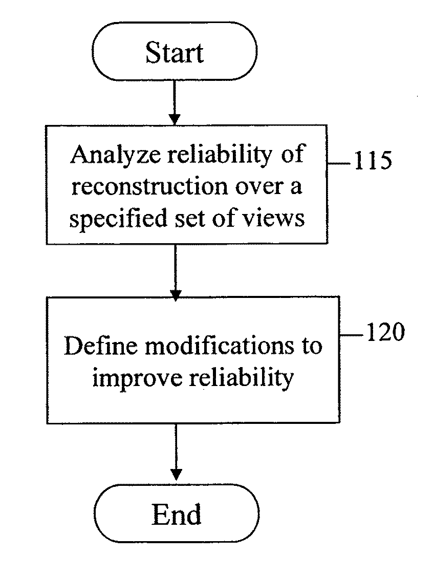
[0099]The present embodiments teach providing modifications of the reconstruction and / or imaging processes in order to obtain a reliable reconstruction of the imaged volume. Specifically, the methods teach analyzing the reliability of the reconstruction obtainable from collected emission data over a set of views to determine modifications which are expected to improve reliability. The present embodiments further teach using radioactive-emission measurements to define views for further radioactive-emission measurements of a body structure, to be performed during the current measurement process.

[0100]With non-uniform scanning, the amount of information available for different voxels is not uniform, thereby constraining the ability to obtain an accurate reconstruction of the volume. In addition, there may be a need to focus on a region or regions of interest, and to control reconstruction resolution and accuracy where and when necessary. In such cases it is important to provide feature...


PUM

PropertyMeasurementUnit
volumeaaaaaaaaaa
volumeaaaaaaaaaa
radioactive-emission density distributionaaaaaaaaaa

Abstract

A method for stabilizing the reconstruction of an imaged volume is presented. The method includes the steps of performing an analysis of the reliability of reconstruction of a radioactive-emission density distribution of the volume from radiation detected over a specified set of views, and defining modifications to the reconstruction process and / or data collection process to improve the reliability of reconstruction, in accordance with the analysis.

Description

RELATED APPLICATIONS[0001]This application is a National Phase of PCT Patent Application No. PCT / IL2006 / 000840 having International Filing Date of Jul. 19, 2006, which claims the benefit of the following U.S. Provisional Patent Applications: 60 / 816,970 filed on Jun. 28, 2006, 60 / 800,846 filed on May 17, 2006, 60 / 800,845 filed on May 17, 2006, 60 / 799,688 filed on May 11, 2006, 60 / 763,458 filed on Jan. 31, 2006, 60 / 750,597 filed on Dec. 15, 2005, 60 / 750,334 filed on Dec. 15, 2005, 60 / 750,287 filed on Dec. 13, 2005, 60 / 741,440 filed on Dec. 2, 2005, 60 / 720,652 filed on Sep. 27, 2005, 60 / 720,541 filed on Sep. 27, 2005, 60 / 720,034 filed on Sep. 26, 2005, 60 / 702,979 filed on Jul. 28, 2005, 60 / 700,753 filed on Jul. 20, 2005, 60 / 700,752 filed on Jul. 20, 2005, 60 / 700,318 filed on Jul. 19, 2005, 60 / 700,299 filed on Jul. 19, 2005 and 60 / 700,317 filed on Jul. 19, 2005.[0002]PCT Patent Application No. PCT / IL2006 / 000840 is also a Continuation-in-Part of the following PCT Patent Applications: PCT...

Claims


Application Information

Patent Timeline
Patent Type & Authority Patents(United States)
IPC IPC(8): G06K9/00
CPCA61B5/02755A61B5/415A61B5/418A61K51/02A61K51/04A61K51/0402A61K51/0476A61K51/0478A61K51/048A61K51/0489A61K51/0491A61K51/1203A61B5/06
Inventor ROUSSO, BENNYDICHTERMAN, ELIZIV, OMERBEN-HAIM, SHLOMO
Owner SPECTRUM DYNAMICS MEDICAL LTD
Why Eureka
Try Eureka