Medical X-ray examination apparatus and method for k-edge imaging

a k-edge imaging and x-ray technology, applied in the field of medical x-ray examination apparatus, can solve the problems general inconvenient use of monochromatic sources in clinical applications, and achieve the effect of high count-rate capabilities

Active Publication Date: 2012-07-24
KONINK PHILIPS ELECTRONICS NV
View PDF12 Cites 4 Cited by
  • Summary
  • Abstract
  • Description
  • Claims
  • Application Information

AI Technical Summary

Benefits of technology

[0006]It is an object of the present invention to provide a medical X-ray examination apparatus and a corresponding method for performing k-edge imaging of an object of interest including material showing k-edge absorption which allows the use of conventional detector technology, which does not suffer from the above described limitation to provide very high count-rate capabilities.
[0015]The invention is based on the idea to use a conventional (polychromatic) X-ray source and a conventional X-ray detector for acquiring at least two sets of projection data. These projection data are acquired using radiation transmitted through a Bragg filter at different Bragg reflection angles. For instance, in a preferred embodiment, a first projection data set is acquired at a certain projection angle at a first Bragg reflection angle and a second projection data set is acquired (preferably at the same or substantially the same projection angle) at a second Bragg reflection angle. From the obtained projection data the desired k-edge image can be reconstructed. Such k-edge images are very desirable and allow, for instance, a direct measurement of a contrast agent such as Gd (or other contrast materials also showing a k-edge effect) injected into a patient.
[0025]This embodiment has the advantage that it is not required that two projection data sets are obtained for at different Bragg reflection angles at the same or substantially the same projection angle. Further, no measures need to be taken for correction of imaging errors as with the above described embodiment.
[0029]In a preferred embodiment the Bragg filter is a Highly Oriented Pyrolytic Graphite (HOPG) filter. Such a HOPG-filter has a certain spectral width and improves the sensitivity.
[0036]According to this aspect of the present invention the two projection data sets obtained at the two different Bragg reflection angles can be acquired simultaneously. Thus, the advantage of requiring more hardware elements compared to the apparatus according to the above explained first aspect is that the data acquisition requires less time.
[0039]Preferably, the X-ray examination apparatus according to the present invention is used for the direct measurement of a contrast medium, such as a contrast agent used in medical imaging. This opens a number of new clinical features to CT imaging such as absolute blood volume measurement or cerebral perfusion imaging. It can enhance the contrast for angiography and allow the discrimination of the contrast agent filled lumen and calcified plaque within a vessel. Preferred contrast agents contain, for instance, iodine or, even more preferred due to a k-edge effect at a higher energy, gadolinium. The invention can further be applied in molecular imaging to reconstruct images showing a special substance, such as a special contrast agent, injected into a patient which only docks to certain cells or other targets, such as tumor cells or fibrin. The method according to the invention thus helps or can be used for quantitative measurements of such cells within a region of interest.

Problems solved by technology

However, monochromatic sources are generally not suitable for clinical applications since they either have power levels far away from the required power for medical imaging or since they use synchrotron radiation of high energy accelerators.
The major roadblock towards spectral CT, however, is the availability of detectors with very high count-rate capabilities.

Method used


Image

  • Medical X-ray examination apparatus and method for k-edge imaging
  • Medical X-ray examination apparatus and method for k-edge imaging
  • Medical X-ray examination apparatus and method for k-edge imaging

Examples


Embodiment Construction

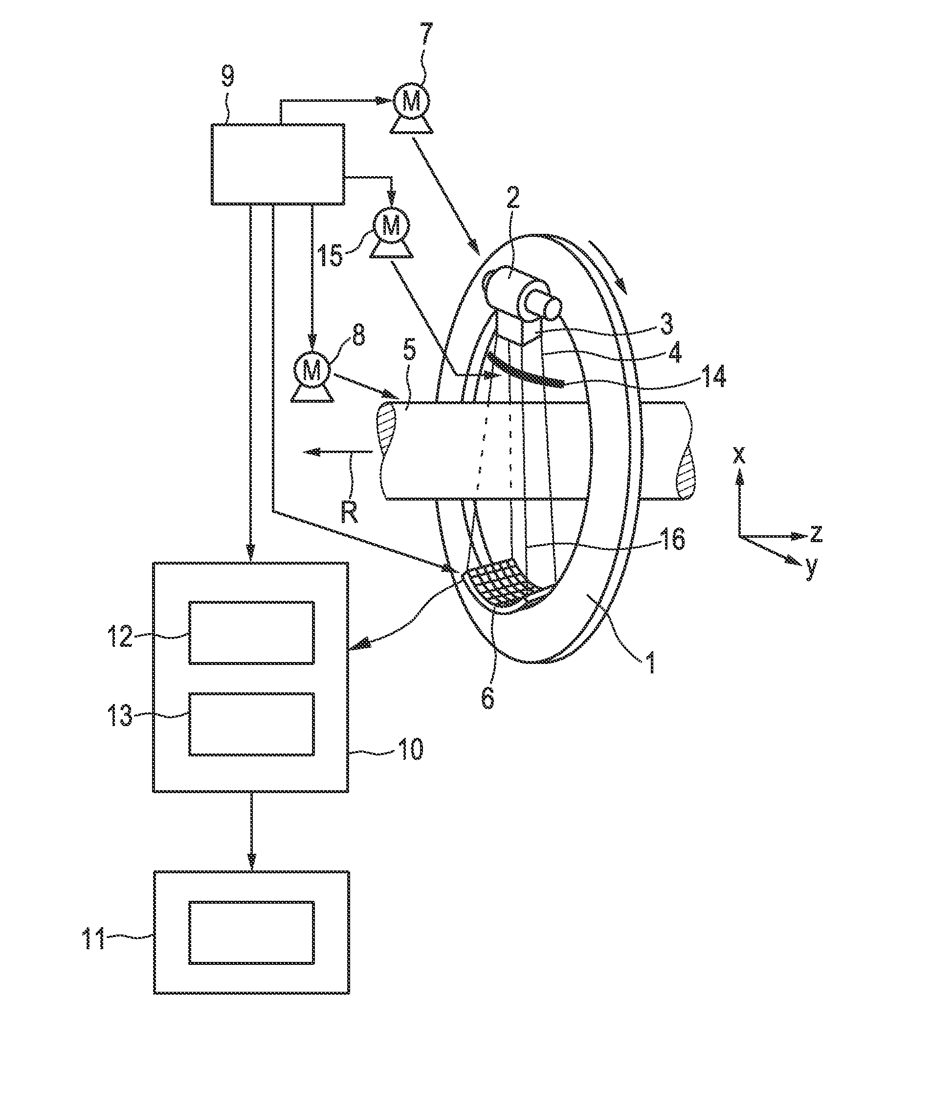
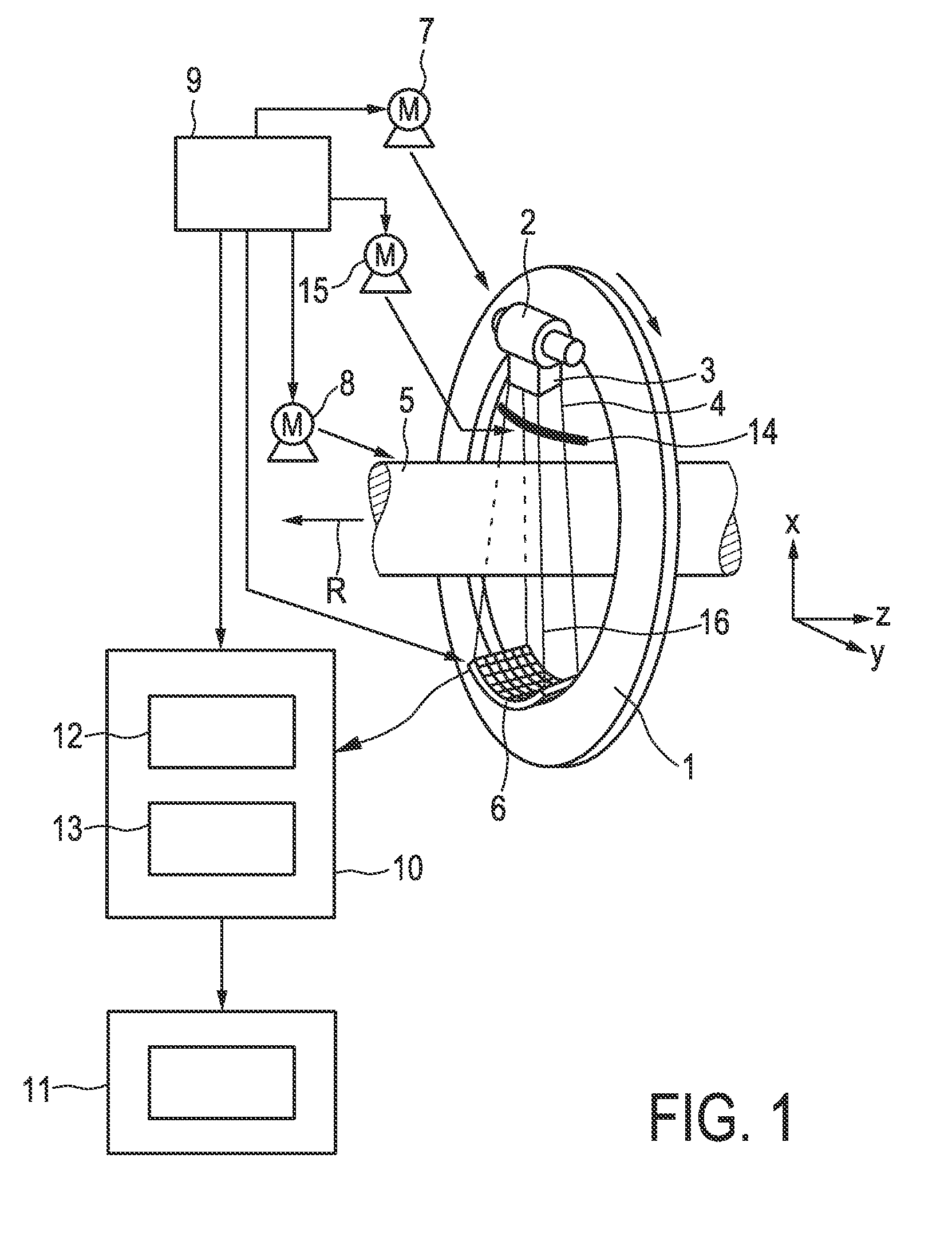
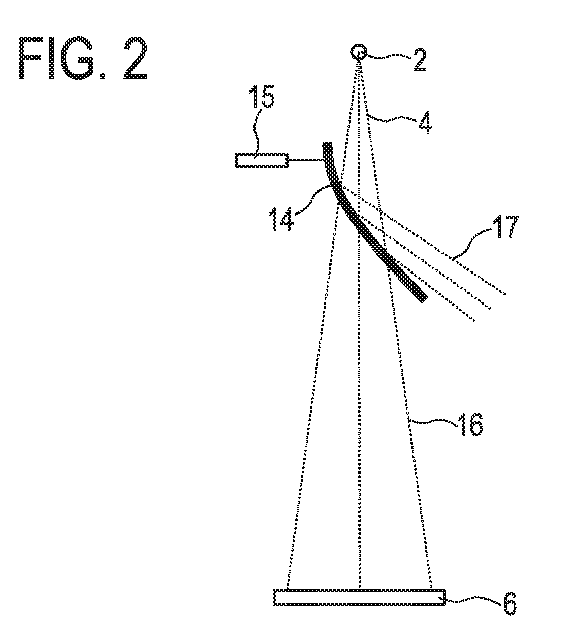
[0048]FIG. 1 shows a first embodiment of a medical X-ray examination apparatus according to the present invention, in particular a CT imaging system. The CT imaging system shown in FIG. 1 includes a gantry 1 which is capable of rotation about an axis of rotation R which extends parallel to the z direction. The radiation source 2, in particular a (conventional) polychromatic X-ray tube for emitting a broad energy spectrum of X-rays, is mounted on the gantry 1. The X-ray tube 2 is provided with a collimator device 3 which forms a conical radiation beam 4 from the radiation produced by the X-ray tube 2. The radiation traverses an object (not shown), such as a patient, in a region of interest in a cylindrical examination zone 5. After having traversed the examination zone 5, the X-ray beam 4 is incident on an X-ray detector unit 6, in this embodiment a two-dimensional detector, which is mounted on the gantry 1.

[0049]The gantry 1 is driven at a preferably constant but adjustable angular ...


PUM


Abstract

The present invention relates to a medical X-ray examination apparatus and method for performing k-edge imaging of an object of interest including material showing k-edge absorption. To allow the use of conventional detector technology, which does not suffer from the limitation to provide very high k-rate capabilities a method is proposed comprising the steps of: —emitting polychromatic X-ray radiation (4; 4a, 4b), —Bragg filtering said polychromatic X-ray radiation by a Bragg filter such that radiation (16) transmitted through said Bragg filter (14; 14a, 14b) passes through said object (5), —detecting X-ray radiation after passing through said object (5), —acquiring projection data at at least two different Bragg reflection angles of said Bragg filter (14; 14a, 14b), and —reconstructing a k-edge image from the acquired projection data.

Description

FIELD OF THE INVENTION[0001]The present invention relates to a medical X-ray examination apparatus and a corresponding method for performing k-edge imaging of an object of interest including material showing k-edge absorption.BACKGROUND OF THE INVENTION[0002]Conventional CT (Computed Tomography) imaging systems measure the X-ray attenuation and provide limited contrast for medical imaging. Most clinical applications use contrast agents to enhance the contrast. However, it would be desired to extend the information contents of CT imaging systems.[0003]In k-edge imaging a tuneable, monochromatic source is used for detection of specific atoms by measuring the attenuation at two or more energies, generally before and behind the k-edge, which is, for instance, described in H. Elleaune, A. M. Charvet, S. Corde, F. Esteve and J. F. Le Bas, “Performance of computed tomography for contrast agent concentration measurements with monochromatic X-ray beams: comparison of k-edge versus temporal s...

Claims


Application Information

Patent Timeline
Patent Type & Authority Patents(United States)
IPC IPC(8): A61B6/00G06K9/00
CPCA61B6/032A61B6/4014A61B6/4035A61B6/4042A61B6/4241A61B6/583A61B6/482A61B6/504A61B6/481G21K2201/062G21K2201/064A61B6/507
Inventor PROKSA, ROLAND
Owner KONINK PHILIPS ELECTRONICS NV
Why Eureka
Try Eureka