System for analyzing tissue perfusion using concentration of indocyanine green in blood

a tissue perfusion and concentration technology, applied in the field of systems for analyzing tissue perfusion and a method of measuring the perfusion rate, can solve the problems of unsuitable method of measuring and currently impossible to clinically measure the precise rate of tissue perfusion using this method

Inactive Publication Date: 2012-10-09
KOREA ADVANCED INST OF SCI & TECH
View PDF94 Cites 33 Cited by
  • Summary
  • Abstract
  • Description
  • Claims
  • Application Information

AI Technical Summary

Problems solved by technology

The conventional tissue perfusion measuring method ‘laser Doppler imaging’ is a method of measuring the degree of scattering of laser light depending on the speed of blood flow in a skin surface, but has a shortcoming in that it is unsuitable for a method of measuring variation in the state in which blood flow is low because sensitivity is low when blood flow rate decreases to less than 20% of the normal of the blood flow rate.
Accordingly, it is currently impossible to clinically measure the precise rate of tissue perfusion using this method.

Method used


Image

  • System for analyzing tissue perfusion using concentration of indocyanine green in blood
  • System for analyzing tissue perfusion using concentration of indocyanine green in blood
  • System for analyzing tissue perfusion using concentration of indocyanine green in blood

Examples


example 1

Establishment of Method of Measuring Perfusion Using ICG

[0071]The present inventors derived an equation through the following steps so as to establish a method of measuring perfusion based on the ICG concentration dynamics in blood.

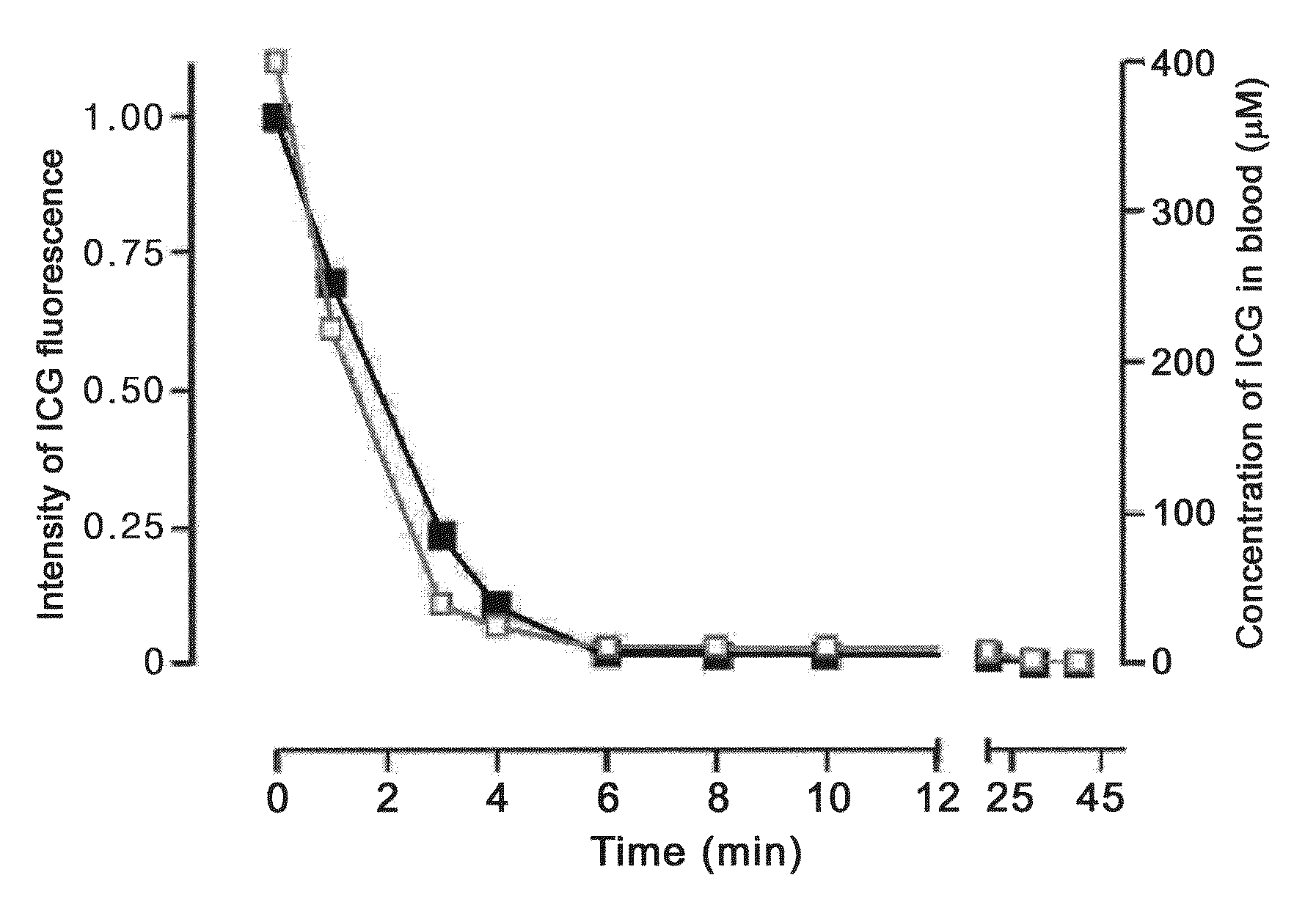
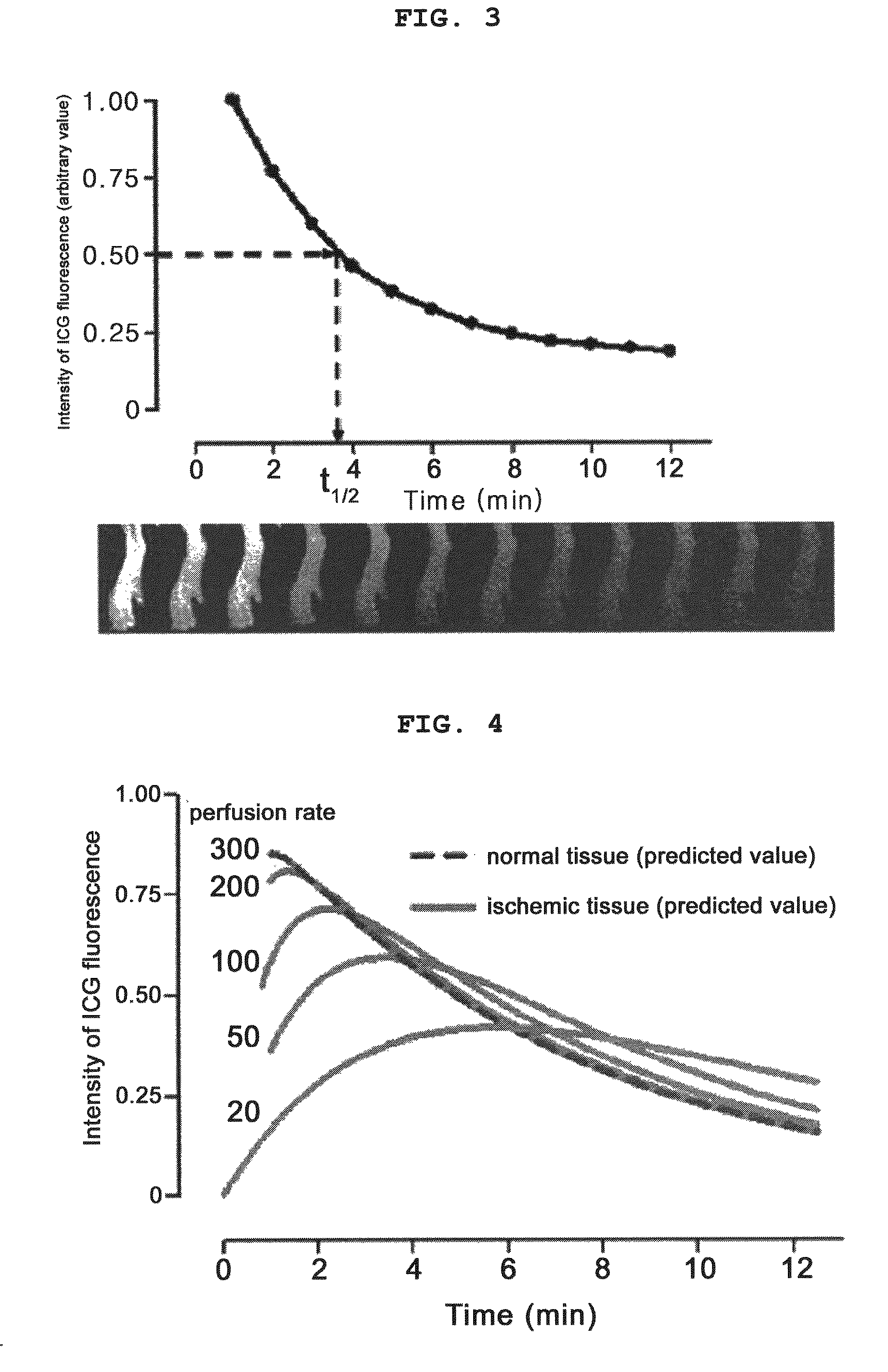
[0072]FIG. 9 is a graph that is obtained by injecting ICG (1.5 mg / kg, Sigma, USA) into a vein, collecting blood over time, measuring the intensities of ICG fluorescence, and converting the intensities into the concentrations of ICG in blood. The closed squares of the graph indicate the intensities of ICG fluorescence, while the open squares thereof indicate the concentrations of ICG in blood. FIG. 3 shows variation in ICG fluorescence over a period of time from 1 minute to 12 minutes after the injection of ICG. That is, in FIG. 3, an image of the ICG fluorescence of a living body acquired with the passage of time is converted into numerical values, with the highest brightness set to 1, and is represented in the form of a graph. When variation in ICG in no...

example 2

Measurement of Perfusion Using Indocyanine Green and the Construction of Perfusion Map and Tissue Necrosis Probability Map Based on Correlation Coefficient

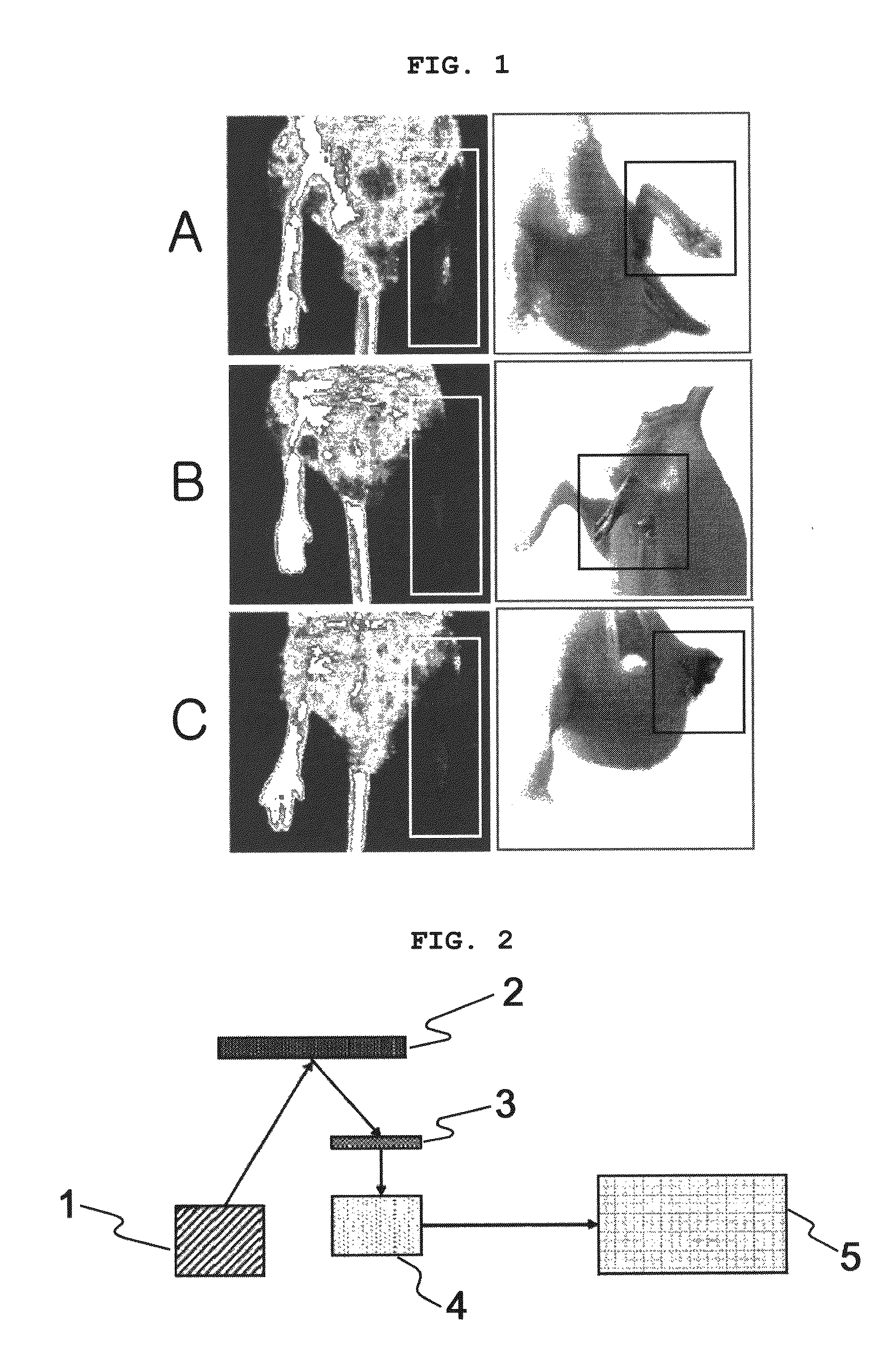
[0084]The present inventors constructed a blood perfusion reduction model to obtain ICG images in an actual ischemic tissue. A surgical operation of removing an artery and vein from one leg of each of 50 nude mice (Charlse liver Japan, Inc.) was carried out, ICG images were acquired using the measurement apparatus of FIG. 2 four hours after the surgical operation, and the perfusion rates of tissues were obtained using the above-described method.

[0085]The obtained perfusion rates of tissues were arranged in a perfusion map (the left side of FIG. 8). In FIG. 8, a perfusion rate of 300% / min corresponds to 9 in the perfusion map, 0% / min corresponds to 0.

[0086]Since the rate of tissue necrosis is considerably affected by the rate of tissue perfusion, whether a ‘perfusion rate’ has been precisely measured can be evaluated through the ob...


PUM


Abstract

The present invention relates, in general, to a system for analyzing tissue perfusion using the concentration of indocyanine green and a method of measuring the perfusion rate using the system and, more particularly, to a system for measuring tissue perfusion by injecting indocyanine green into a living body, detecting variation in the concentration of indocyanine green with the passage of time, and analyzing the detected variation, and a method of measuring the perfusion rate using the system. The present invention provides a method of measuring perfusion in a living body, which enables accurate measurement for respective regions in a wide range from a perfusion rate decreased to less than 10% of normal perfusion to a perfusion rate increased to greater than normal perfusion using the above-described mechanism of ICG in a living body, which cannot be conducted using the conventional technology.

Description

RELATED APPLICATIONS[0001]This application is a 35 U.S.C. §371 national stage filing of International Application No. PCT / KR2007 / 003355 filed on Jul. 11, 2007, which claims priority to, and the benefit of, Korean Patent Application No. 10-2006-0099033 filed on Oct. 11, 2006. The contents of the aforementioned applications are hereby incorporated by reference.TECHNICAL FIELD[0002]The present invention relates to a system for analyzing tissue perfusion and a method of measuring the perfusion rate using the system. More particularly, the present invention relates to a system for measuring tissue perfusion by injecting indocyanine green into a living body, detecting variation in the concentration of indocyanine green with the passage of time, and analyzing the detected variation, and a method of measuring the perfusion rate using the system.BACKGROUND[0003]The conventional tissue perfusion measuring method ‘laser Doppler imaging’ is a method of measuring the degree of scattering of lase...

Claims


Application Information

Patent Timeline
Patent Type & Authority Patents(United States)
IPC IPC(8): A61B5/00
CPCA61B5/0059A61B5/0261A61B5/0275A61B6/00A61B5/00
Inventor CHOI, CHULHEEKANG, YUJUNGCHOI, MYUNGHWAN
Owner KOREA ADVANCED INST OF SCI & TECH
Why Eureka
Try Eureka