Binding component

a technology of components and components, applied in the field of binding components, can solve the problems of unwieldy and sometimes difficult to manage in the operative field, sharp chiselled points, and difficulty in operative management, and achieve the effect of improving the binding structur

Inactive Publication Date: 2012-11-13
ECOLE DE TECH SUPERIEURE
View PDF10 Cites 31 Cited by
  • Summary
  • Abstract
  • Description
  • Claims
  • Application Information

AI Technical Summary

Benefits of technology

[0064]More specifically, advantages of the present invention include that the proposed binding structure advantageously applies a substantially constant compressive force across tissue boundary while being able to accommodate some expansion.
[0074]Also, the proposed binding structure is designed so as to be manufacturable using conventional forms of manufacturing so as to provide a device that will be economically feasible.

Problems solved by technology

Despite their widespread use, the stainless steel wires suffer from numerous drawbacks that can cause problems both to the surgeon and to the patient during the operation and to the patient after closure is completed.
For example, the relatively stiff and unyielding characteristic of a stainless steel wire renders it unwieldy and sometimes difficult to manage on the operative field.
This substantial problem is compounded by the fact that the wire is typically cut using a wire cutter with relatively blunt blades, which generates a chiselled point that is typically quite sharp.
However, such clamps are also plagued with various drawbacks including cluttering of the operating field and being tedious and time consuming to work with and around.
Furthermore, the stainless steel sternal wires can disrupt the entire image generated in a computerized axial tomography or magnetic resonance imaging scan of the chest for the remainder of the patient's life.
Still furthermore, tightening by twisting wires together with a pair of pliers is an inexact method.
Consequently, some suture wires break during installation.
Sternal wires occasionally also break after the surgery.
Such breakage can be secondary to the thinning and deformation of the steel strand by the excessive force or stresses that are sometimes applied to the loop during routine closure.
For fear of breaking a wire, a surgeon may tend to undertorque the suture, resulting in less than optimal closure pressure on the sternal knit line.
This, in turn, can lead to dehiscence problems.
A particularly major problem associated with steel wire sutures is that post-operative stress on the closure loops may cause the thin wires to cut into and through the bone of the sternum.
Indeed, since wires inherently define a relatively small contact surface, anatomical structures may experience excessive localized pressure resulting in damage.
The coughing, deep breathing and movement required to attain these goals imposes substantial stresses on the sternal closure.
Elderly patients or patients who have thin or osteoporotic bones are particularly susceptible to this complication.
The result is further loosening of the sternal closure which can lead to painful instability of the two sternal halves with respiratory compromise and ultimately sternal dehiscence.
Instability of the sternal closure can also result in internal bleeding.
This, in turn, can increase the risks of infection and / or result in macerative damage to the cartilage and associated muscle tissue with a consequent increase in post-operative discomfort and in the time required for healing.
Also, if a second operation for sternal rewiring is required, it is made even more difficult by the fact that the sternal halves are often sliced into pieces by the stainless steel wires.
However, these proposed solutions tend to result in increased damage to blood vessels or other soft tissue, and also may substantially increase the time required for closing the chest.
Also, if infection occurs necessitating removal of the sutures, it can be very difficult to remove the reinforcing wires.
However, they nevertheless suffer from various limitations which limit their utility.
For example, being substantially flat and made of relatively stiff and unyielding material, they are typically unable to fittingly contact the geometry of the sternum.
Also, their geometry is such that they cannot penetrate easily through the bone and, hence, can only be positioned peristernally between the ribs.
Consequently, they are considered unergonomical.
Typically, they are even more unwieldy and difficult to manage on the operative field than steel wires.
Still furthermore, the substantially flat shape of these bands results in relatively sharp side edges.
This, in turn, may cause internal haemorrhaging and associated problems.
The sharp side edges, if unprotected, also have considerable potential to slide into the fingers of the operating surgeon or assistants.
Furthermore, they are capable of inflicting injury to the soft tissues below the sternum during closure.
However, the components thereof are relatively large and may cause serious pain or other ailments to the patient.
A common problem to all of the hereinabove mentioned bone binding structures is that they can only be used towards fixation of the sternal halves, i.e. for immobilizing the sternal halves in close proximity to each other.
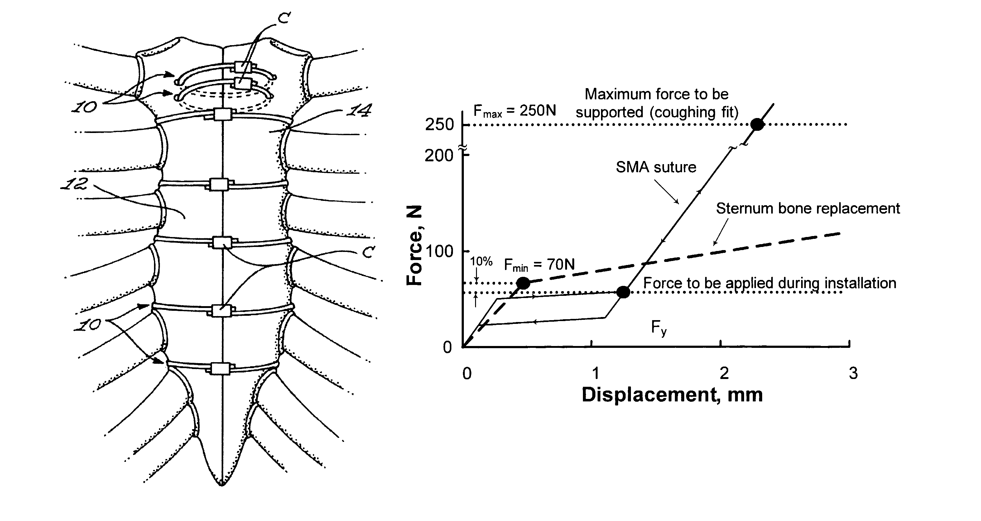
The above-mentioned structures cannot respond to this dimensional change and, consequently, cannot maintain compression across the facing boundaries of the divided sternum during the healing process.
Thus, applied pressure decreases with time.
As stated above, not only do the large initial compression forces generated with the hereinabove mentioned devices diminish in the initial phase of bone healing, but such large forces, in themselves, are detrimental relative to the concentrated forces experienced proximal to the wires.
Although the hereinabove mentioned structures provide some stability, they are deficient as a means to establish a known initial force and they never reconcile the need for continuous compressive force.
Furthermore, physiological activities such as coughing contribute to the degeneration not only of the sternum but also potentially of the devices themselves.
With increased deformation, there is generally a work-hardening effect, in which the increased tangle of dislocations makes additional deformation more difficult.
Binding devices disclosed in the prior art using shape-memory alloys typically suffer from numerous drawbacks.
For example, the structure disclosed in U.S. Pat. No. 5,766,218 is relatively complex to manufacture and, hence, potentially less reliable and more expensive.
Furthermore, the use of a strap is associated with the hereinabove mentioned disadvantages inherent to its geometry.
However, again, they suffer from disadvantages inherently associated with their geometries.

Method used


Image

  • Binding component
  • Binding component
  • Binding component

Examples


example 1

[0143]As seen from FIG. 7, the superelastic behaviour of the shape memory alloy (SMA) allows a non-zero zero force to be applied to the sternum even though the width of the sternum tends to decrease as a result of the nature of the healing process. This decrease in the width of the sternum is represented by portions A-B-C of the stress-strain relationship illustrated in FIG. 7. In addition, SMA benefit from the dynamic interference phenomenon [9], and manifest significant hardening under external impulses (for example coughing), as seen in the C-D portion of the stress-strain curve illustrated in FIG. 7, thus providing a quasi-constant pressure onto the sternum once a predetermined load has been reached, as seen from the D-E portion of the curve of FIG. 7. This quasi-constant pressure is defined by the height of the upper plateau of the superelastic loop. Once the disruption is over, the force applied by the closure system to the sternum returns to its initial level on the lower pla...

experimental study

[0161]To consider the geometry of the SMA sternum closure system, a series of experimental tests were undertaken.

Description of the Testing Bench

[0162]FIG. 11 shows the outline of the testing bench used. Two identical closure systems (7) were installed simultaneously on polyurethane blocks (2) simulating the sternum. The installation force was applied with the help of two adjustable loading clamps (3). Two LC703-100 load cells (5) (Omega, Stamford, Conn., USA) installed on each extremity of the testing frame (1) allowed the force at the sternum interface to be measured. An Enduratec ELF 3200 tensile testing machine (4) was used to apply external force. LabView 6.0 (National Instruments Corp., Austin, Tex., USA) data acquisition systems registered the real time displacement of the testing machine's piston as well as the forces measured by the load cells.

Components Used

[0163]A comparison was made between two closure devices: (1) Ø0.78 mm No 5 Ethicon steel wire and Ø3 mm SMA braided 1...


PUM


Abstract

A binding component for binding together a pair of biological tissues. The binding component includes an elongated body defining a body longitudinal axis, the body being made, at least in part, of a shape memory material. The body is configured and sized so as to be both substantially flexible and substantially compressible in a direction substantially perpendicular to said longitudinal axis.

Description

[0001]This is a National Stage Application of PCT / CA05 / 01859 under 35 USC §371(a), which claims priority of U.S. Provisional Patent Application Ser. No. 60 / 633,512 filed Dec. 6, 2004.FIELD OF THE INVENTION[0002]The present invention relates to the general field of binding components and is particularly concerned with a binding component suitable for binding together a pair of biological tissues.BACKGROUND OF THE INVENTION[0003]There exists a plurality of situations wherein it is desirable to bind together components having specific properties. For example, there are various circumstances in which separated tissue of a patient needs to be brought together so it can heal. Such tissue may include bone, muscle, fascia or the like that has been divided to gain access for example to the thoracic cavity, the mediastinum, the abdomen or the like.[0004]Typically, most surgical procedures involving the heart or lungs are performed through a midline sternal incision, widely referred to as medi...

Claims


Application Information

Patent Timeline
Patent Type & Authority Patents(United States)
IPC IPC(8): A61B17/03A61B17/82
CPCA61B17/0487A61B17/06166A61B17/82A61B17/823A61B2017/00867
Inventor BRAILOVSKI, VLADIMIRCARTIER, RAYMONDTERRIAULT, PATRICKBARIL, YANNICK
Owner ECOLE DE TECH SUPERIEURE
Why Eureka
Try Eureka