
Medical imaging system for segementing blood vessel

a medical imaging and blood vessel technology, applied in the field of medical imaging systems, can solve the problems of manual segmentation, reduced efficiency of clinicians, slow manual segmentation, and prone to human errors

Inactive Publication Date: 2013-07-16
SIEMENS HEALTHCARE GMBH
View PDF4 Cites 7 Cited by
  • Summary
  • Abstract
  • Description
  • Claims
  • Application Information

AI Technical Summary

Problems solved by technology

Such manual segmentations have reduced efficiency of clinicians, mis-detection of blood vessels, etc.
Manual segmentation is slow and prone to human errors, thus automatic segmentation of the blood vessels is desired.

Method used


Image

  • Medical imaging system for segementing blood vessel
  • Medical imaging system for segementing blood vessel
  • Medical imaging system for segementing blood vessel

Examples


Embodiment Construction

[0012]The embodiments herein and the various features and advantageous details thereof are explained more fully with reference to the non-limiting embodiments that are illustrated in the accompanying drawings and detailed in the following description. Descriptions of well-known components and processing techniques are omitted so as to not unnecessarily obscure the embodiments herein. The examples used herein are intended merely to facilitate an understanding of ways in which the embodiments herein may be practiced and to further enable those of skill in the art to practice the embodiments herein. Accordingly, the examples should not be construed as limiting the scope of the embodiments herein.

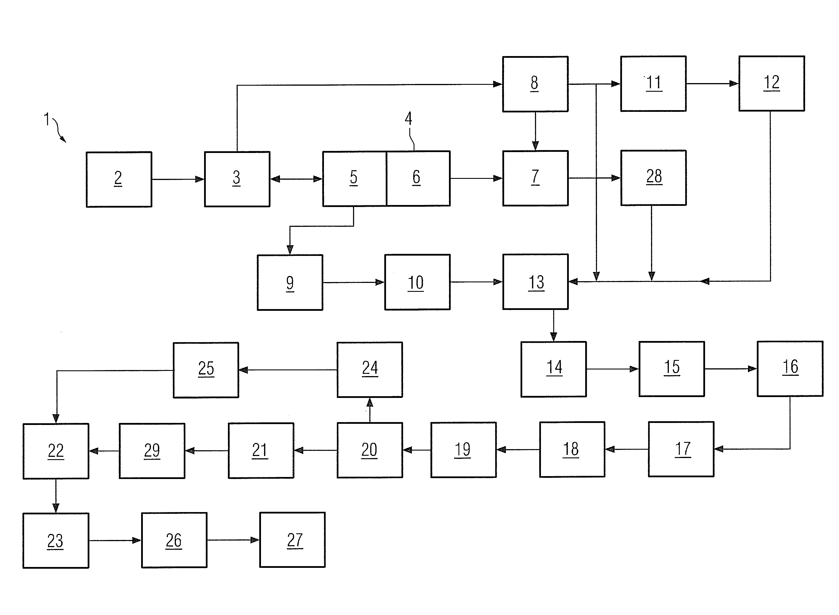
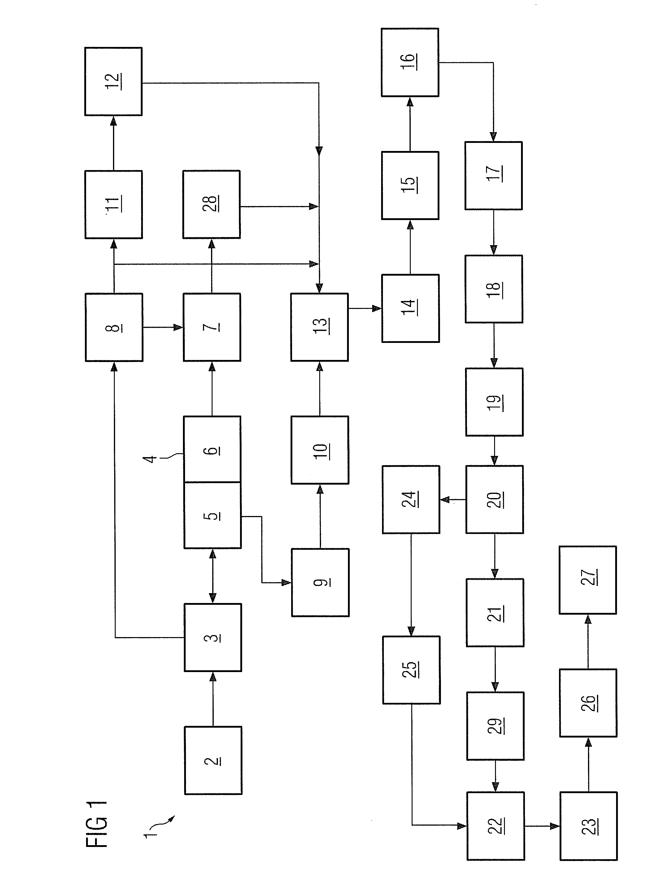
[0013]FIG. 1 illustrates a schematic diagram of a system 1 to segment an original blood vessel of a body part represented by an original medical image 2 in accordance with an embodiment herein. As illustrated, the system 1 includes an image analyzer 3 which receives the original medical image 2...


PUM


Abstract

A medical imaging system to segment an original blood vessel of a body part represented by an original medical image is provided. The system includes an image analyzer for receiving and the original medical image to analyze the original medical image to provide a Hessian Eigen analysis comprising a first data and a second data mapped to each pixel of the medical image, and an image identifier for receiving the Hessian Eigen analysis and for identifying seed points from the pixels by processing the first data and the second data along with a vesselness property, wherein the seed points are used for segmenting the original blood vessel to provide a corrected medical image representing a corrected blood vessel.

Description

FIELD OF INVENTION[0001]The embodiments herein generally relate to medical imaging systems, and, more particularly, to a medical imaging system for segmenting blood vessels on a basis of saptio-temporal metric from electro-cardiograph data input.BACKGROUND OF INVENTION[0002]Medical images represent various parts of the human body like blood vessels, bones, etc. By analyzing the medical images clinicians can plan treatment for the patients and at the same time, the clinicians can be guided to operate the body part while operating over the body part, for example catheter guidance, image guidance for the surgery. The medical images for surgery guidance are analyzed by registering the medical image with pre-operative 3D medical data. During the registration, the blood vessels represented by the medical images are used as features, which further requires segmentation of the blood vessels in real-time. Currently, segmentation of the blood vessels is done manually by clinicians which invol...

Claims


Application Information

Patent Timeline
Patent Type & Authority Patents(United States)
IPC IPC(8): G06K9/00
CPCG06T7/0081G06T7/0093G06K2209/057G06T2207/20148G06T2207/20156G06T2207/30101G06T7/11G06T7/162G06T7/136G06V2201/034
Inventor GUPTA, VIPINKALE, AMITSUNDAR, HARI
Owner SIEMENS HEALTHCARE GMBH
Learn More