Tibial augments for use with knee joint prostheses
a knee joint and tibial technology, applied in the field of tibial augmentation for use with knee joint prostheses, can solve the problems of removing an unnecessary amount of healthy bone, adding additional tibia parts, and other problems, and achieving the effect of improving the quality of life and reducing the risk of fractur
- Summary
- Abstract
- Description
- Claims
- Application Information
AI Technical Summary
Benefits of technology
Problems solved by technology
Method used
Image
Examples
first embodiment
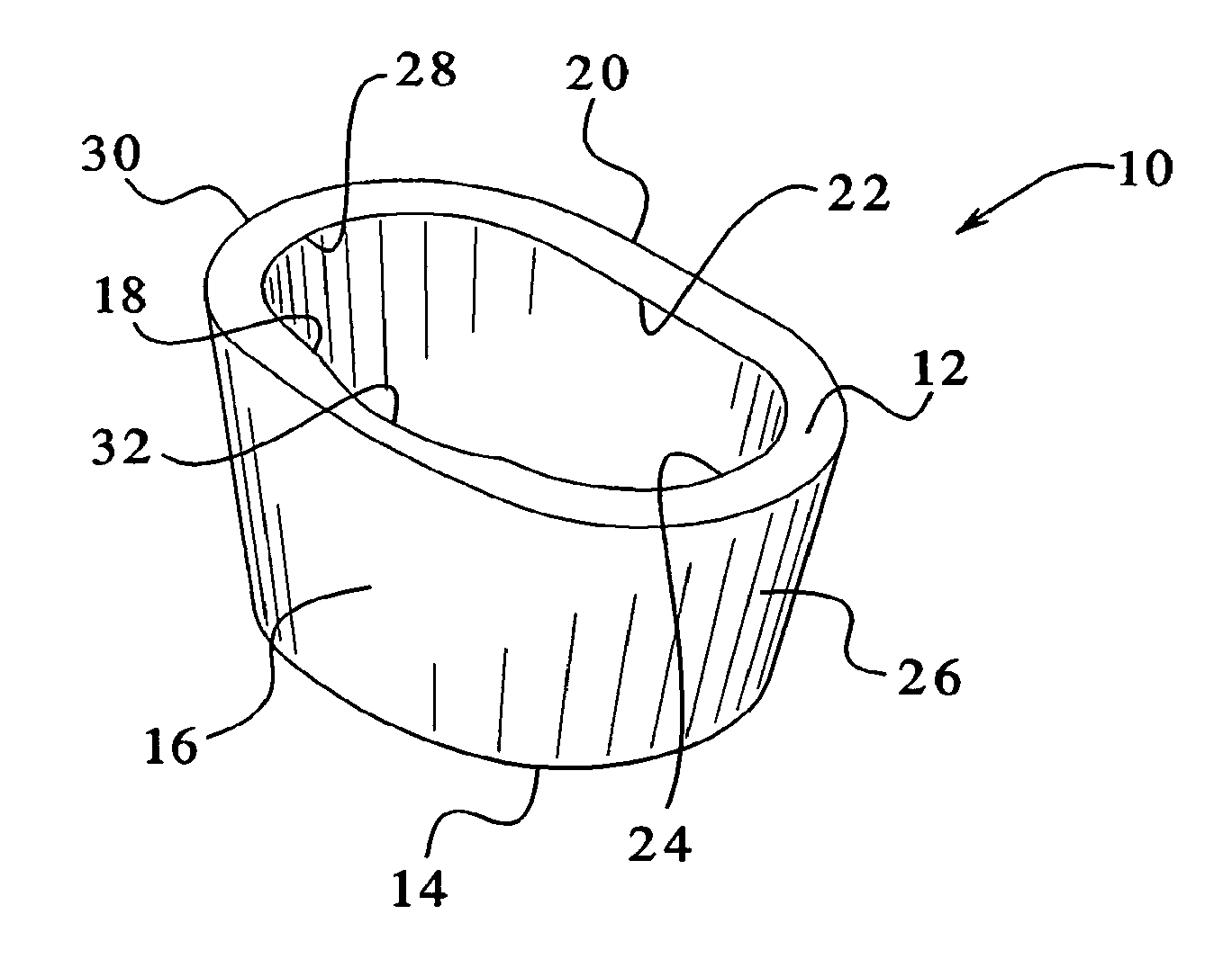
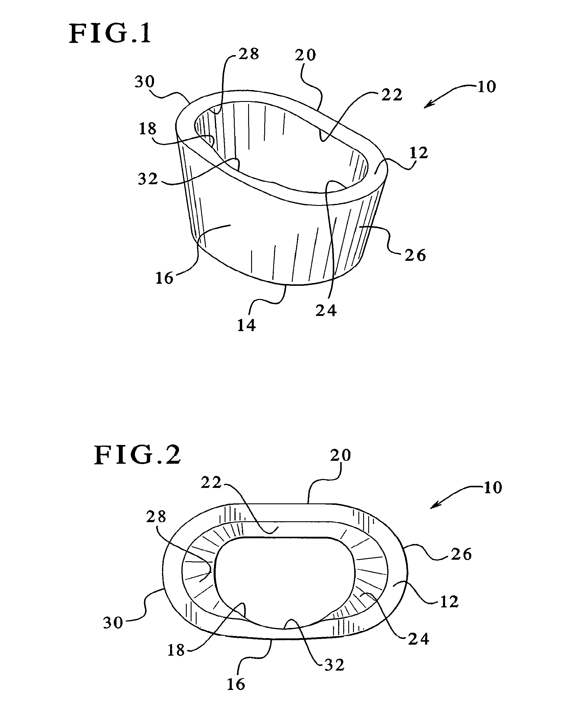
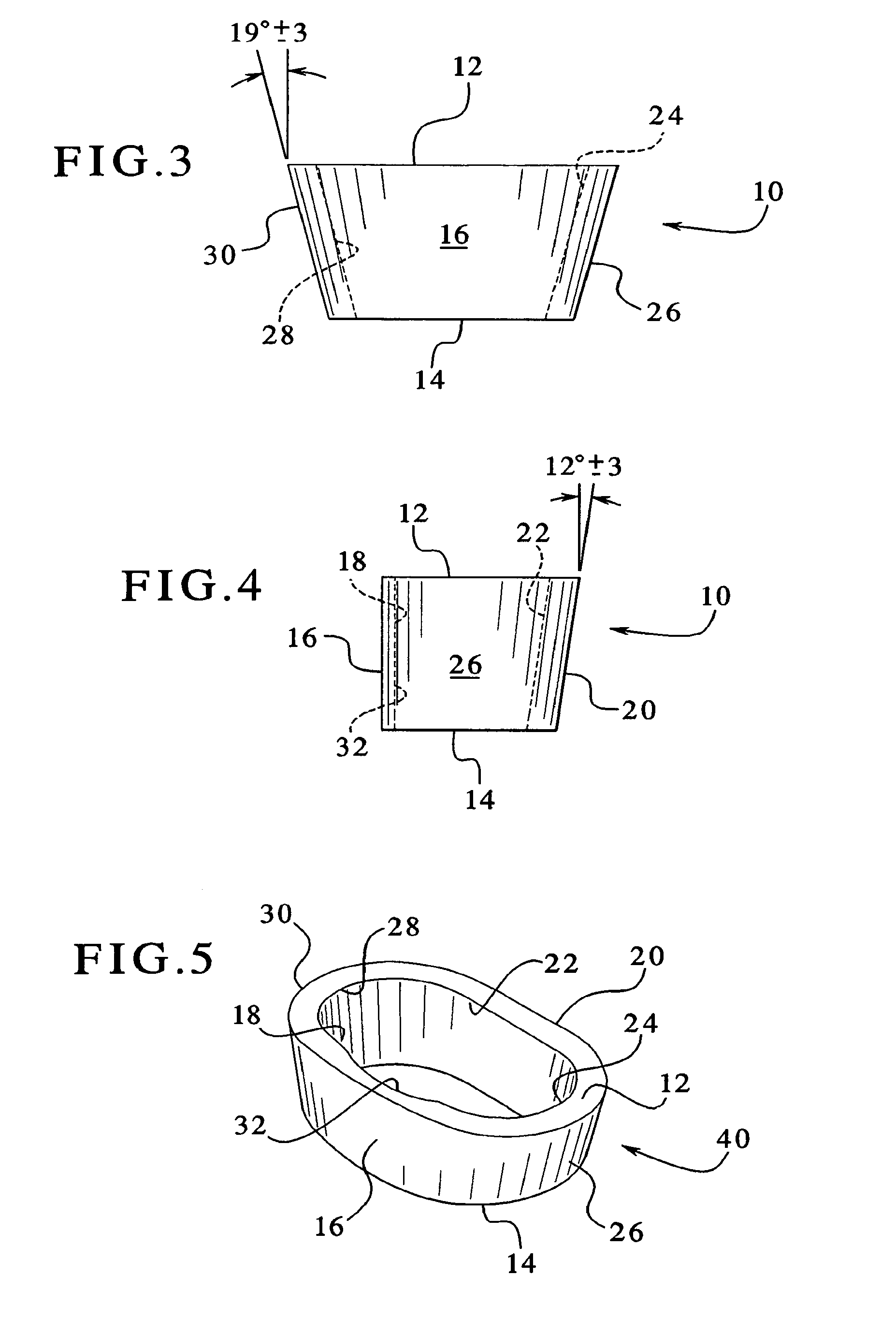
[0042]Referring to FIGS. 1 through 4, the tibial augment of the present invention is shown. The tibial augment is preferably an annular member 10, and it is preferably made from a tantalum based porous material, such as Trabecular Metal™. Trabecular Meta1™ is desirable because it resembles bone and approximates the physical and mechanical properties of bone better than other materials. Use of such a metal enables increased bonding with the adjacent bone by allowing the bone to grow into its highly porous surface. The tibial augment may also be made of other materials, and it is preferably made of a material that facilitates bony ingrowth.
[0043]The tibial augment 10 is anatomically sized and shaped to fill an existing cavitary defect within the proximal human tibia or a cavity prepared in the proximal portion of a human tibia. In the preferred embodiment, a system of different stock sizes of augments would be available, as discussed more fully below, with different sizes being used f...
second embodiment
[0057]a stepped tibial augment is shown in FIG. 6B, as represented by tibial augment 60. In this embodiment, as in the FIG. 6A embodiment, distal surface 14a is located at a greater distance from the proximal surface 12 than distal surface 14b. The main difference between this embodiment and the FIG. 6A embodiment is the location of the transition surface 52. In this embodiment, the transition surface 52 is located closer to the outer lateral surface 26 than to the outer medial surface 30.
third embodiment
[0058]FIG. 6C shows a stepped tibial augment 70. In this embodiment, as in the embodiments of FIGS. 6A and 6B, distal surface 14a is located at a greater distance from the proximal surface 12 than distal surface 14b. The main difference between this embodiment and the other two embodiments is the location of the transition surface 52. In this embodiment, the transition surface 52 is located closer to the outer medial surface 30 than to the outer lateral surface 26.
[0059]The embodiments of FIGS. 6A through 6C are especially useful where to there has been uneven tibial decay, i.e., where there is more decay on either the lateral side or the medial side than on the other side. By using one of the stepped tibial augments shown in FIGS. 6A through 6C, more healthy bone, if it exists on one side or the other, can be preserved, and mostly only defective bone will end up being removed when forming a stepped cavity to implant the tibial augment. In other words, the base of the cavity into wh...
PUM
 Abstract
Description
Claims
Application Information
 - R&D
- Intellectual Property
- Life Sciences
- Materials
- Tech Scout
- Unparalleled Data Quality
- Higher Quality Content
- 60% Fewer Hallucinations
Browse by: Latest US Patents, China's latest patents, Technical Efficacy Thesaurus, Application Domain, Technology Topic, Popular Technical Reports.
© 2025 PatSnap. All rights reserved.Legal|Privacy policy|Modern Slavery Act Transparency Statement|Sitemap|About US| Contact US: help@patsnap.com


