Tissue anchor system

a technology of tissue anchoring and tissue, which is applied in the direction of staples, nails, infusion needles, etc., can solve the problems of increasing the complexity of the device structure, raising particular challenges, and challenging the use of percutaneous technology,

Active Publication Date: 2015-01-27
KARDIUM
View PDF516 Cites 8 Cited by
  • Summary
  • Abstract
  • Description
  • Claims
  • Application Information

AI Technical Summary

Benefits of technology

[0042]The implant kit may further include a plurality of fasteners, each fastener movable along a respective one of the guide lines to a position where at least some of the fasteners secure the implant member to the tissue under tension in the deployed configuration. Each of the fasteners may include a unidirectional clutch that in use allows the fastener to advance along a respective one of the guide lines toward a respective one of the embedded tissue anchors and prevents the fastener from retreating along the guide line away from the respective embedded tissue anchor. The plurality of fasteners and the implant member may be provided in a unitary structure. The at least some of the fasteners may each be fastenable to a respective one of the guide lines to secure the implant member to the tissue under tension in the deployed configuration. The at least some of the fasteners may each be fastenable to a respective one of the embedded tissue anchors to secure the implant member to the tissue under tension in the deployed configuration.

Problems solved by technology

However, the use of percutaneous technologies also raises some particular challenges.
Medical devices used in percutaneous surgery need to be deployed via narrow tubes called catheter sheaths, which significantly increase the complexity of the device structure.
As well, doctors do not have direct visual contact with the medical tools used once they are placed within the body, and positioning the tools correctly and operating the tools successfully can often be very challenging.
Improper function of any one of these structures, alone or in combination can lead to mitral regurgitation.
The mechanical valve carries the risk of thromboembolism and requires anticoagulation with all of its potential hazards, whereas the biological prosthesis suffers from limited durability.
Another hazard with replacement is the risk of endocarditis.
Such a procedure is expensive, requires considerable time, and is associated with high morbidity and mortality.
Due to the risks associated with this procedure, many of the sickest patients are denied the potential benefits of surgical correction of mitral regurgitation.
In addition, patients with moderate, symptomatic mitral regurgitation are denied early intervention and undergo surgical correction only after the development of cardiac dysfunction.
Furthermore, the effectiveness of such procedures is difficult to assess during the procedure and may not be known until a much later time.
Hence, the ability to make adjustments to or changes in the prosthesis function to obtain optimum effectiveness is extremely limited.
Both approaches suffer from several problems as a result of attempting to reshape the mitral annulus using an alternative method.
Devices that deform the coronary sinus, while suitable for percutaneous procedures, are not effective in controlling the leakage of the mitral valve as the forces are not applied from the correct opposite sides of the valve, which are the lateral annulus and the septal annulus.
The devices of the second type are not easily adapted to a percutaneous procedure.
Pulling applied along a different direction will distort the mitral valve but will not achieve the optimal approximation of the two leaflets.

Method used


Image

  • Tissue anchor system
  • Tissue anchor system
  • Tissue anchor system

Examples


Embodiment Construction

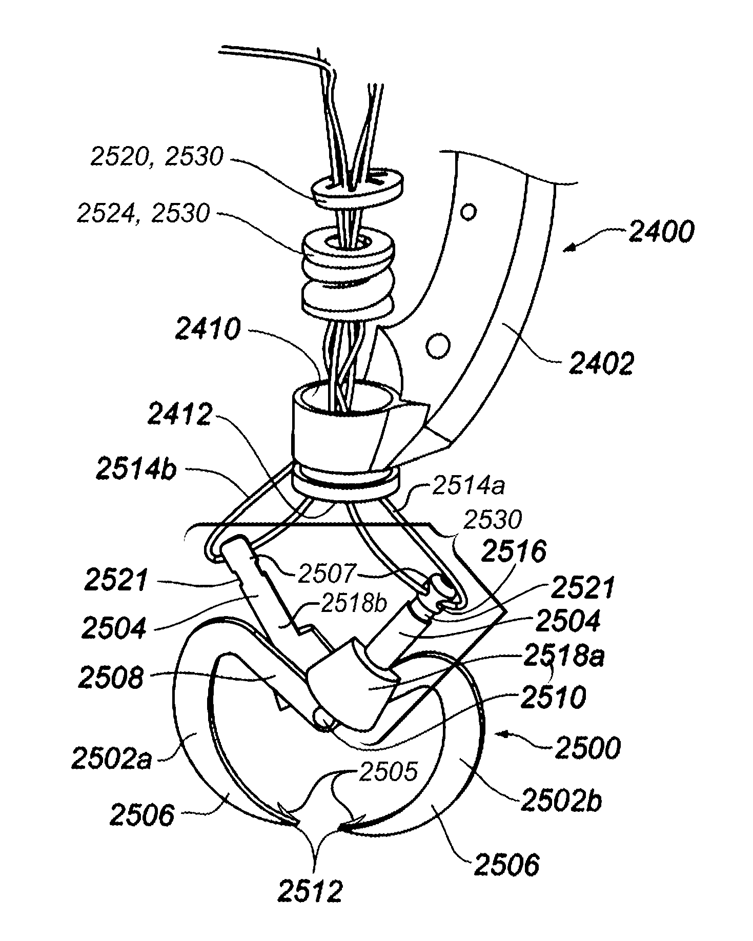
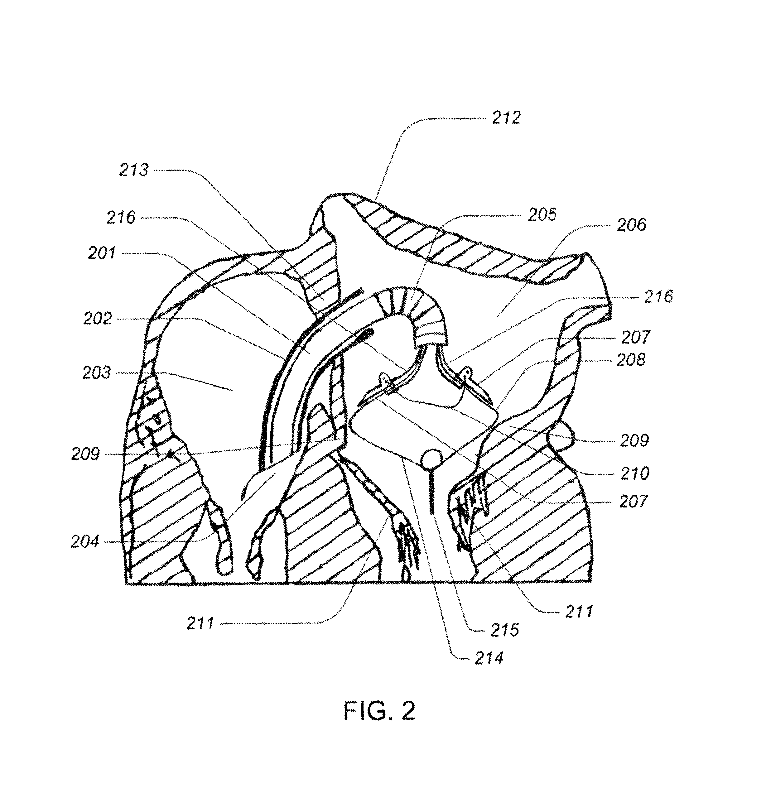
[0114]In the following description, certain specific details are set forth in order to provide a thorough understanding of various embodiments of the invention. However, one skilled in the art will understand that the invention may be practiced without these details. In other instances, well-known structures have not been shown or described in detail to avoid unnecessarily obscuring descriptions of the embodiments of the invention.

[0115]Reference throughout this specification to “one embodiment” or “an embodiment” means that a particular feature, structure or characteristic described in connection with the embodiment is included in at least one embodiment of the present invention. Thus, the appearances of the phrases “in one embodiment” or “in an embodiment” in various places throughout this specification are not necessarily all referring to the same embodiment. Furthermore, the particular features, structures, or characteristics may be combined in any suitable manner in one or more...


PUM


Abstract

A tissue anchor system includes a plurality of elongated members pivotally coupled by a pivot member and at least one coupler coupled to at least one of the elongated members between a pivot point and an end of the elongated member which coupler is operable to secure the elongated members in a configuration that securely couples the tissue anchor to tissue.

Description

CROSS-REFERENCE TO RELATED APPLICATIONS[0001]This application claims benefit under 37 U.S.C. 119(e) to U.S. patent application Ser. No. 61 / 388,405, filed Sep. 30, 2010, which is incorporated by reference herein in its entirety.BACKGROUND[0002]1. Field[0003]This disclosure is generally related to percutaneous or minimally invasive surgery, and more particularly to tissue anchor systems for use in percutaneously or minimally invasive surgery.[0004]2. Description of the Related Art[0005]Cardiac surgery was initially undertaken only by performing a sternotomy, a type of incision in the center of the chest, which separates the sternum (chest bone) to allow access to the heart. In the previous several decades, more and more cardiac operations are performed using a percutaneous technique, which is a medical procedure where access to inner organs or other tissue is gained via a catheter.[0006]Percutaneous surgeries benefit patients by reducing surgery risk, complications, and recovery time....

Claims


Application Information

Patent Timeline
Patent Type & Authority Patents(United States)
IPC IPC(8): A61B17/08A61B17/04A61B17/064A61B17/068A61F2/24
CPCA61B17/064A61B17/0401A61B17/068A61F2/2448A61F2/2466A61B17/0644A61B2017/0409A61B2017/0414A61B2017/0437A61B2017/0441A61B2017/0464A61B2017/0645A61B2017/0647A61B2017/0649
Inventor GOERTZEN, DOUGLAS
Owner KARDIUM
Why Eureka
Try Eureka