In situ tissue engineering using magnetically guided three dimensional cell patterning

a three-dimensional cell patterning and in situ tissue technology, applied in the field of in situ tissue engineering using magnetically guided three-dimensional cell patterning, can solve the problems of not being able to predictably achieve functional hyaline cartilage, inability to transfer approaches to clinical settings, laborious procedures,

Active Publication Date: 2015-06-09
SCRIPPS HEALTH
View PDF0 Cites 1 Cited by
  • Summary
  • Abstract
  • Description
  • Claims
  • Application Information

AI Technical Summary

Benefits of technology

[0006]The approach presented herein is a means to circumvent the deficiencies in the field of regenerative medicine generally, and allows for the production of organized tissues in situ with specific cellular organizations that mimic the native tissue. The creation of organized repair tissue may provide more appropriate three-dimensional cues for tissue formation. Direct implantation and organization in situ may circumvent the need for long-term in vitro culture, and may also allow for more complex tissue types to be formed. The present invention addresses this and other needs.
[0007]The present invention provides an approach for manipulation of cells in a three dimensional manner with the use of magnetic particles. The methods provided herein allow for the control of cell distance, cell density, and cell orientation within a single hydrogel without specialized “pre-functionalization” of the magnetic particles.

Problems solved by technology

However, this procedure does not predictably result in functional hyaline cartilage, but rather in fibrocartilage (Roberts et al.
Although in vitro prefabrication of tissue grafts has shown variable success in animal models and cartilage tissues engineered with zonal organization has improved to provide a natural spatial cell distribution, these approaches have not been transferred to a clinical setting (Chu et al.
The procedures are laborious, expensive and integration into the surrounding host cartilage tissue is an unresolved issue (Obradovic et al.

Method used


Image

  • In situ tissue engineering using magnetically guided three dimensional cell patterning
  • In situ tissue engineering using magnetically guided three dimensional cell patterning
  • In situ tissue engineering using magnetically guided three dimensional cell patterning

Examples


example 1

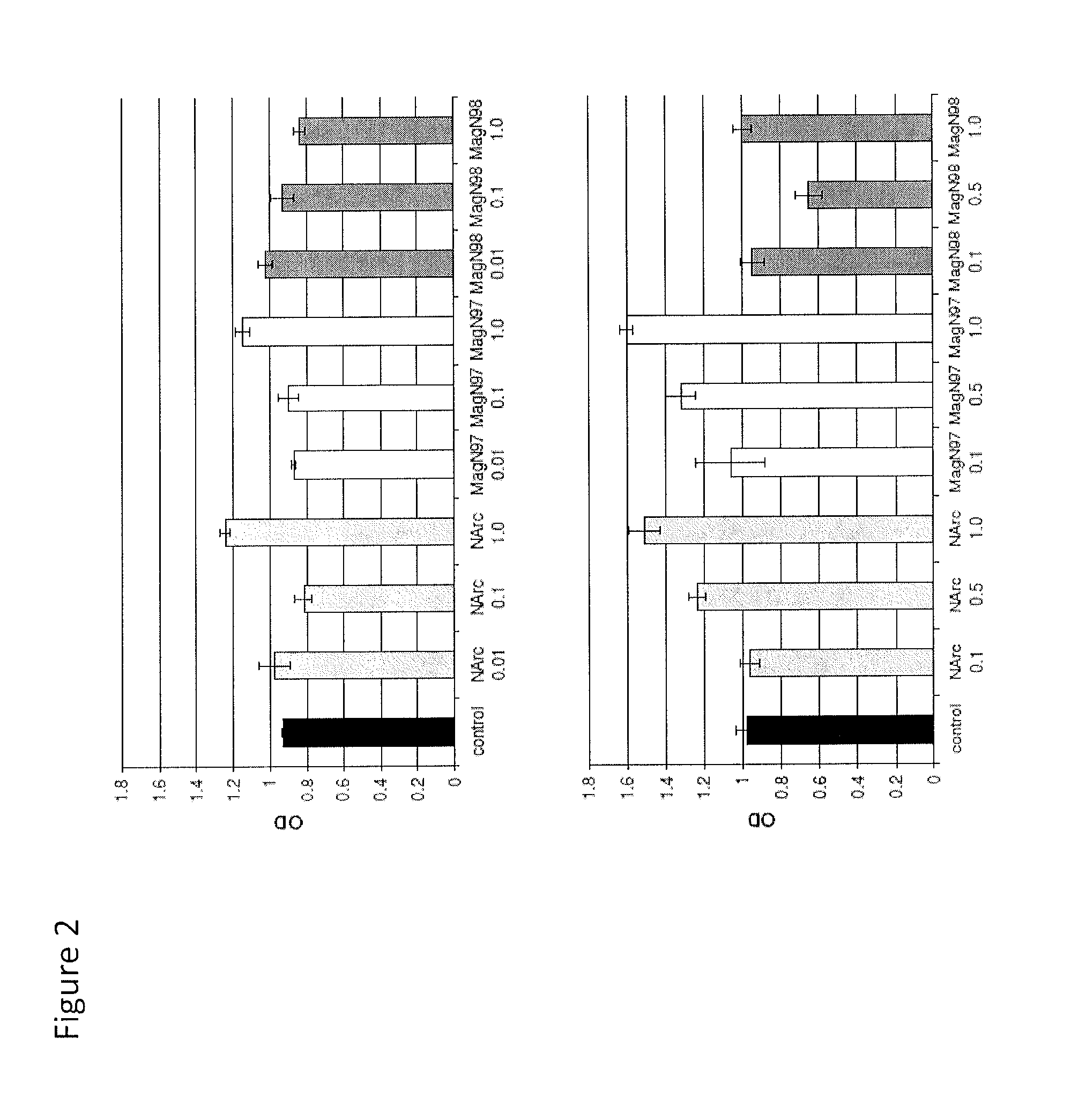
Mammalian Cell Viability in Response to Iron Oxide Particles

[0145]Three iron oxide materials were examined:

[0146](i) NanoArc Industrial maghemite (Fe2O3), 20-40 nm diameter, “NArc”;

[0147](ii) Magnetite (Fe3O4) 97%-325 mesh, ˜44 μm diameter, “MagN97” and

[0148](iii) Magnetite (Fe3O4) 98% 20-30 nm diameter (MagN98).

[0149]The iron oxide materials were obtained from the same source (Alfa Aesar, Ward Hill, Mass.). Each particle was weighed and washed in 5 mL absolute ethanol once, centrifuged for 5 minutes at 2000 rpm, washed with 1×PBS three times (5 mL), and finally resuspended in PBS at a weight to volume of 10 mg / mL. Each mixture was sterilized via autoclave.

[0150]Human articular cartilage was obtained from tissue banks (see above). Chondrocytes were isolated via enzymatic digestion and cultured for one passage.

[0151]Specifically, human chondrocytes were seeded in 96-well plates (5000 cells per well) and precultured overnight in DMEM with 2% calf serum. Following pre-culture, the cell...

example 2

Evaluation of Gene Expression in Human Chondrocytes Mixed With Iron Oxide Particles

[0164]Human chondrocyte pellets cultures (5×10⁵ cells each) were formed in the presence of iron oxide particles (1 mg / mL).

[0165]Three species of iron oxide particles were tested separately:

(i) NanoArc industrial maghemite (Fe2O3), 20-40 nm (NArc);

(ii) Magnetite (Fe3O4) 97%-325 mesh (˜44 μm) (MagN97) and

(iii) Magnetite (Fe3O4) 98% 20-30 nm (MagN98).

[0166]Cell pellet cultures were maintained in serum-free ITS+ medium supplemented with TGFβ1 (10 ng / mL), for 12 days (as described by Barber et al. (2004). Osteoarthritis Cartilage 12, pp. 476-484). The medium was changed every 3 days. After 12 days, some pellets were fixed and embedded in paraffin for histology, while other pellets were prepared for RNA extraction for gene expression analysis.

Gene Expression Analysis

[0167]Cells were harvested, and total RNA was isolated from the cultured cell pellets, using the RNAeasy mini kit (Qiagen, Hilden, Germany). Fi...

example 3

Evaluation of Gene Expression in High Density Magnetically-Labeled Chondrocyte-Alginate Cultures

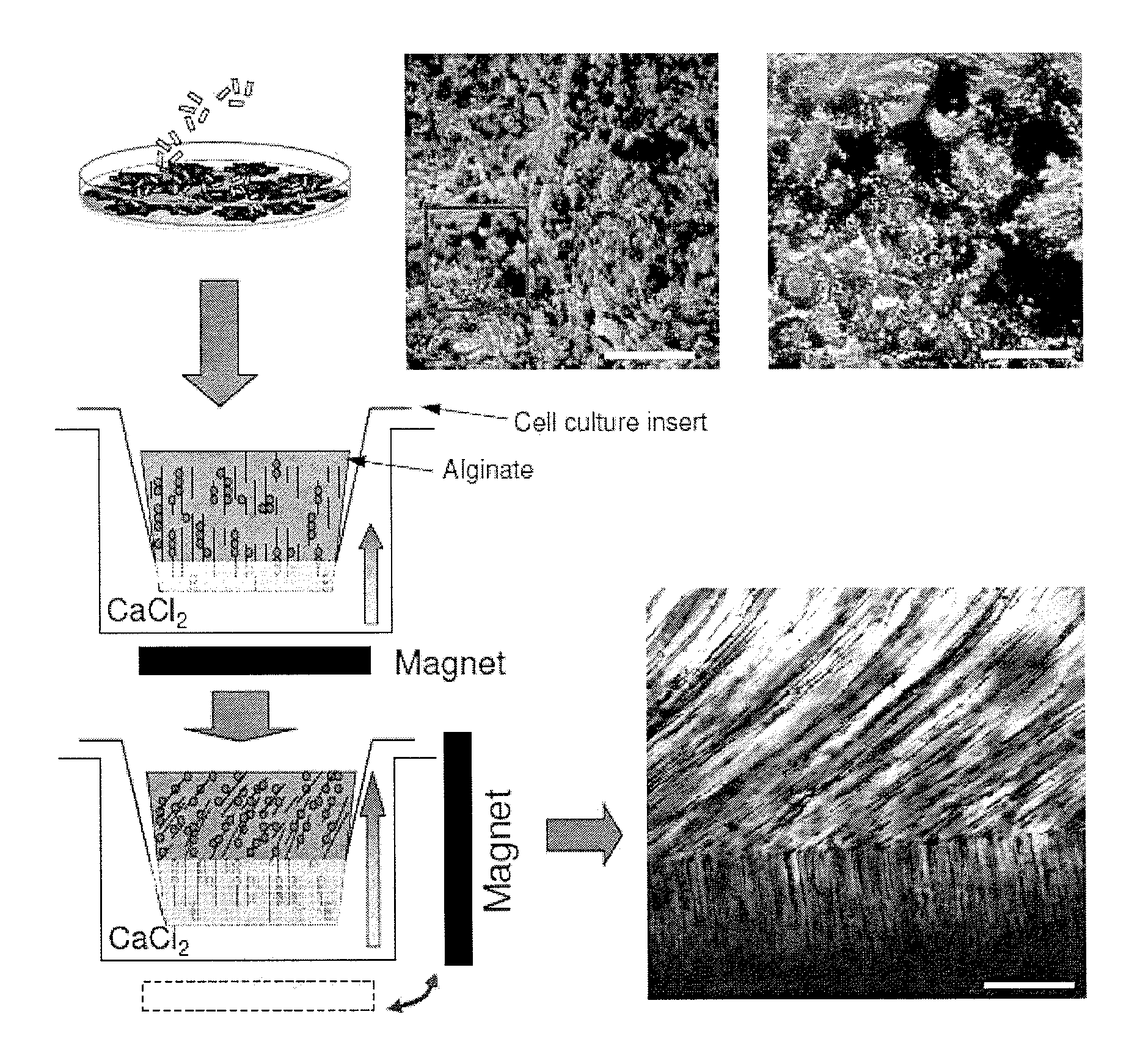
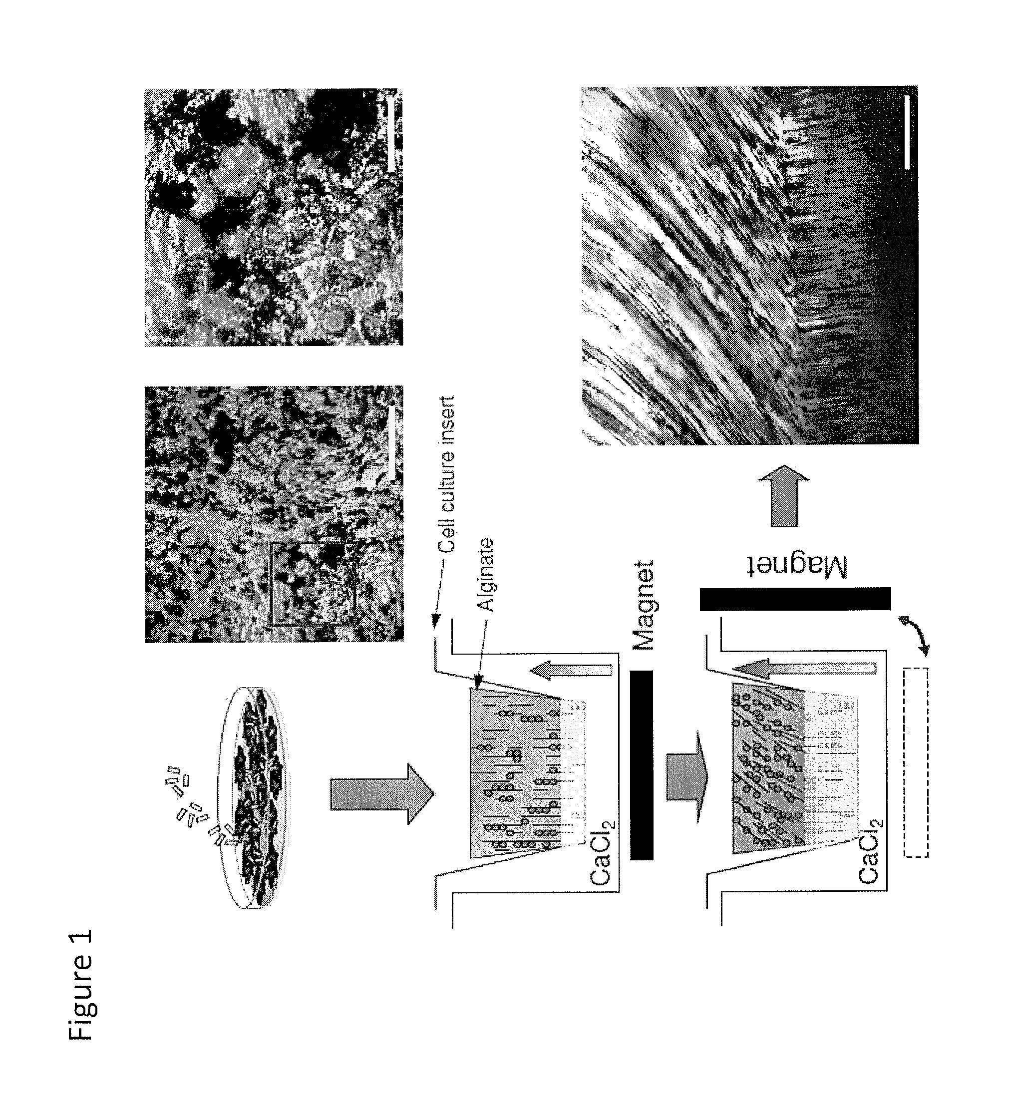
[0174]Human chondrocytes were incubated with MagN97, MagN98 or NArc (1 mg / mL) for 24 hours. Particles rapidly adhered to cells within 30-40 minutes and some particles were engulfed by the cells over 24 hours. Following removal of excess particles by washing in PBS, the cells were detached, mixed into 2% alginate, and transferred into cell-culture inserts, and cultured for two weeks (FIG. 1). The cultures were developed using 2×10⁶ cells.

[0175]Three species of iron oxide particles (1 mg / mL) were tested separately.

Gene Expression Analysis

[0176]Cells were harvested and total RNA was isolated from the isolated cells using the RNAeasy mini kit (Qiagen, Hilden, Germany). First strand cDNA synthesis was performed using total RNA as a template according to the manufacturer's protocols (Applied Biosystems, Foster City, Calif.). Quantitative real time PCR was performed using TagMan® gene expression...


PUM

PropertyMeasurementUnit
magnetic fieldaaaaaaaaaa
diameteraaaaaaaaaa
diameteraaaaaaaaaa

Abstract

Methods are provided for the three dimensional manipulation of cells, and for the formation of an organized engineered cell tissue. Also provided are the organized engineered cell tissues produced by the methods. In one method, a plurality of magnetically labeled cells are mixed with a cross-linkable hydrogel to form a cell-hydrogel mixture, the at least a portion of the plurality of magnetically labeled cells are manipulated with a magnetic field to arrange the magnetically labeled cells into a specific cellular arrangement, and the hydrogel is crosslinked to form the organized engineered cell tissue. The approach presented herein offers a means to circumvent the deficiencies in the field of regenerative medicine, and allows for the production of organized tissues in situ with specific cellular organizations that mimic the native tissue.

Description

CROSS REFERENCE TO RELATED APPLICATION[0001]This application claims priority from U.S. Provisional Application Ser. No. 61 / 445,403, filed Feb. 22, 2011, the disclosure of which is incorporated by reference in its entirety for all purposes.BACKGROUND OF THE INVENTION[0002]Degeneration of tissues such as cartilage is a common health condition that has a significant economic impact and quality of life impact. For example, osteoarthritis (OA) and other rheumatic diseases are among the most common health conditions and are the number one cause of disability in the United States, affecting an estimated 27 million Americans in 2008 (Lawrence et al. (2008). Arthritis Rheum. 58, pp. 26-35). The economic impact of arthritis in the United States is estimated at $128 billion per year, representing more than 2% of the gross domestic product ((CDC), C.f.D.C.a.P. MMWR Morb Mortal Wkly Rep. 56, 4-7 (2007)). Only symptom-modifying therapies are used to treat OA (Roddy et al. (2005). Rheumatology (Ox...

Claims


Application Information

Patent Timeline
Patent Type & Authority Patents(United States)
IPC IPC(8): C12N5/077A61K35/12A61K35/00
CPCC12N5/0655A61K35/12C12N2529/00C12N2533/10C12N2533/74
Inventor GROGAN, SHAWN PATRICKD'LIMA, DARRYL DAVIDCOLWELL, JR., CLIFFORD W.JIN, SUNGHO
Owner SCRIPPS HEALTH
Why Eureka
Try Eureka