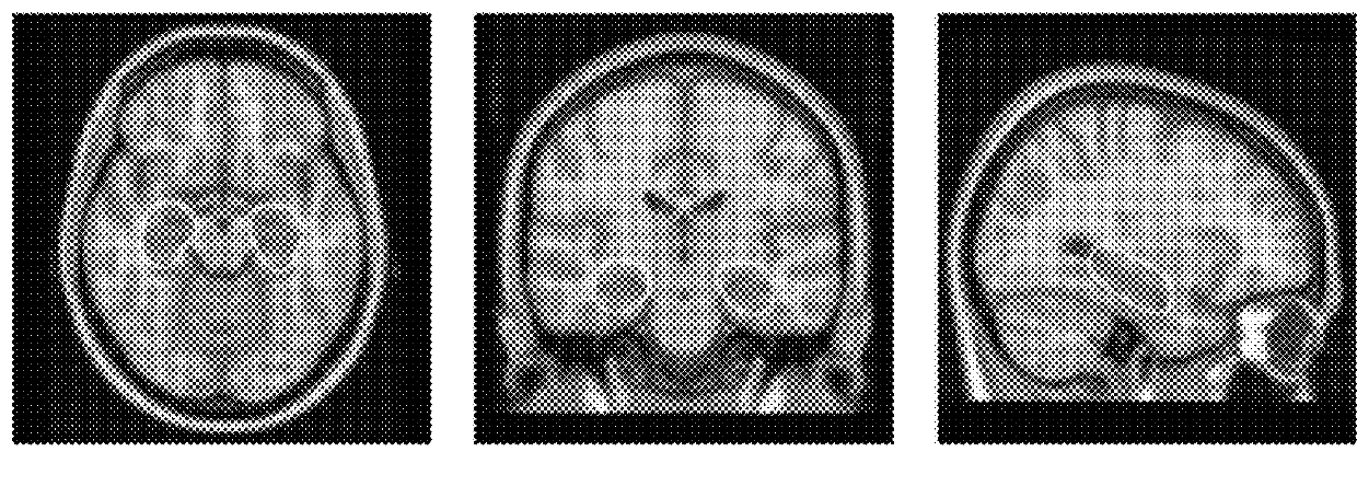
Method and apparatus for processing medical images

a technology for medical images and processing methods, applied in image enhancement, image analysis, instruments, etc., can solve the problems of high-quality manual atlases that are often relatively limited in number, labour-intensive and expensive, and the selection of atlases is difficult, so as to minimize the accumulation of registration errors and improve accuracy

Active Publication Date: 2016-02-02
IP2IPO INNOVATIONS LTD
View PDF6 Cites 6 Cited by
  • Summary
  • Abstract
  • Description
  • Claims
  • Application Information

AI Technical Summary

Benefits of technology

[0014]By virtue of the iterative propagation of label data from the closest labelled images to the unlabelled images, each unlabelled image can be segmented using structurally-similar atlases. As a consequence, relatively large differences between a labelled image and an unlabelled image may be broken down into a number of small differences between comparatively similar initially-unlabelled images through which the label data is propagated, enabling registration errors to be reduced.
[0016]Thus, preferably the step of comparing the labelled and unlabelled images comprises embedding the images into a low-dimensional coordinate system. This enables the labelled and unlabelled images to be compared and the differences to be quantitatively evaluated in a computationally-efficient manner. In certain embodiments the low-dimensional coordinate system may be a two-dimensional coordinate space, thus further simplifying the analysis and processing of the differences between the images.
[0019]Preferably the step of propagating label data comprises propagating label data from a plurality of the closest of the labelled images, based on a classifier fusion technique. This enables the selected image(s) to be labelled with greater accuracy.
[0020]Preferably the method further comprises, after step (d) and before step (e), a step of performing an intensity-based refinement operation on the newly-propagated label data, in order to further minimize the accumulation of registration errors during the labelling process.
[0021]The images may be of different subjects. Alternatively, at least some of the images may be of the same subject but taken at different points in time, thereby enabling intra-subject variance to be identified and studied.

Problems solved by technology

Due to the wide range of anatomical variation, the selection of atlases becomes an important issue in multi-atlas segmentation.
Although an increasing number of MR brain images are available, the generation of high-quality manual atlases is a labour-intensive and expensive task (see e.g. Hammers et al.
This means that atlases are often relatively limited in number and, in most cases, restricted to a particular population (e.g. young, healthy subjects).
This can limit the applicability of the atlas database even if a selection approach is used.
Test data with a greater level of variation than the training data would, however, represent a significant challenge to this approach.
Additionally, the use of a highly variable training dataset may lead to an unrepresentative PCA model as the likelihood of registration errors between the diverse images and the single template is increased.
Prior work where automatically labelled brain images were used to label unseen images did not result in an improvement of segmentation accuracy over direct multi-atlas propagation.

Method used


Image

  • Method and apparatus for processing medical images
  • Method and apparatus for processing medical images
  • Method and apparatus for processing medical images

Examples


Embodiment Construction

[0038]The present embodiments represent the best ways known to the applicants of putting the invention into practice. However, they are not the only ways in which this can be achieved.

[0039]Primarily, the present embodiments take the form of a method or algorithm for processing medical (or other) images. The method or algorithm may be incorporated in a computer program or a set of instruction code capable of being executed by a computer processor. The computer processor may be that of a conventional (sufficiently high performance) computer, or some other image processing apparatus or computer system. Alternatively, the computer processor may be incorporated in, or in communication with, a piece of medical imaging equipment such as an MRI scanner.

[0040]The computer program or set of instruction code may be supplied on a computer-readable medium or data carrier such as a CD-ROM, DVD or solid state memory device. Alternatively, it may be downloadable as a digital signal from a connecte...


PUM


Abstract

The present invention provides, among other things, methods of processing medical images for producing images with labeled anatomical features, including obtaining images containing labeled anatomical features, obtaining unlabelled images, comparing and selecting unlabelled images that most closely resemble labeled images, and propagating label data from labeled images to unlabelled images, thereby labeling corresponding anatomical features on unlabelled images. The present invention also provides systems for performing such methods.

Description

[0001]This invention relates to a method and corresponding apparatus for processing medical images. It is particularly suitable, but by no means limited, for processing magnetic resonance images, for example of the human brain.BACKGROUND TO THE INVENTION[0002]The automated extraction of features from magnetic resonance images (MRI) of the brain is an increasingly important process in neuroimaging. Since the brain anatomy varies significantly across subjects and can undergo significant change, either during aging or through disease progression, finding an appropriate way of dealing with anatomical differences during feature extraction has gained increasing attention in recent years.[0003]Amongst the most popular methods for dealing with this variability are atlas-based approaches. In the context of the present work, an “atlas” is a dataset (which may be a 3D image, a 2D image, images of any dimension, or a set of images) having annotations or labels in order to identify points, regio...

Claims


Application Information

Patent Timeline
Patent Type & Authority Patents(United States)
IPC IPC(8): G06T7/00G06K9/62G06K9/46G06V10/422
CPCG06T7/0089G06K9/468G06K9/6252G06T7/0081G06T2207/10088G06T2207/20128G06T2207/30016G06T7/11G06T7/149G06V10/422G06V10/7715G06F18/21375
Inventor RUECKERT, JAN PAUL DANIELWOLZ, ROBINAJABAR, PAUL
Owner IP2IPO INNOVATIONS LTD
Why Eureka
Try Eureka