Systems and methods for enclosing an anatomical opening

a technology of anatomical opening and anatomical lumen, which is applied in the field of implantable therapeutic devices and endovascular placement of devices, can solve the problems of high risk of anesthesia, bleeding, and infection associated with these types of procedures, and the current surgical approaches available for repairing defects in anatomical lumens and tissues, and repairing defects in septal defects, etc., and other types of anatomical irregularities and defects. high risks

Active Publication Date: 2016-03-08
PULSAR VASCULAR
View PDF255 Cites 19 Cited by
  • Summary
  • Abstract
  • Description
  • Claims
  • Application Information

AI Technical Summary

Benefits of technology

[0048]Several embodiments of the technology are methods and systems directed to reducing the length and complexity of minimally invasive procedures for supporting and occluding openings and repairing a lumen or tissue defect, and to restoring a physiological structure, such as a blood vessel, to its normal, healthy condition. In another aspect, selected embodiments of methods and systems of the present technology provide implantable devices for supporting and / or at least partially occluding and / or at least partially diverting flow away from an opening or cavity, such as an aneurysm, that are safely and conveniently deployable using minimally invasive techniques. Additional features of selected embodiments of the technology may reduce shifting and migration following placement and avoid restricting blood flow in neighboring vessels. In yet another aspect, selected embodiments of methods and systems of the present technology are directed to retaining materials inside a physiological opening or cavity, such as embolic materials within an aneurysm.

Problems solved by technology

Many of the currently available surgical approaches for closing openings and repairing defects in anatomical lumens and tissues (e.g., blood vessels), septal defects, and other types of anatomical irregularities and defects are highly invasive.
Surgical techniques for repairing septal defects are also highly invasive.
The risks related to anesthesia, bleeding, and infection associated with these types of procedures are high, and tissue that is affected during the procedure may or may not survive and continue functioning.
Accurate and timely placement of these mixtures is crucial and very difficult because it is difficult or impossible to control them once they have been placed in the blood flow.
Migration or projection of a vaso-occlusive device from the cavity may interfere with blood flow or nearby physiological structures and poses a serious health risk.
In addition to the difficulties of delivering implantable occlusion devices, some types of aneurysms are challenging to treat because of structural features of the aneurysm or because of particularities of the site.
Wide-neck aneurysms, for example, are known to present particular difficulty in the placement and retention of vaso-occlusive coils.
Aneurysms at sites of vascular bifurcation are another example where the anatomical structure poses challenges to methods and devices that are effective in treating the typical sidewall aneurysms.
In view of such challenges, implanting conventional embolic coils, other structures, or materials in the internal space of an aneurysm has not been an entirely satisfactory surgical approach.
The placement procedure may be arduous and lengthy because it often requires implanting multiple devices, such as coils, serially in the internal space of the aneurysm.
Higher risks of complication from such sources as anesthesia, bleeding, thromboembolic events, procedural stroke, and infection are associated with such longer procedures.
Moreover, because placement of structures in the internal space of an aneurysm does not generally completely occlude the opening, recanalization of the original aneurysm may occur, and debris and occlusive material may escape from within the aneurysm to create a risk of stroke or vessel blockage.
Blood may also flow into the aneurysm and other blood vessel irregularities after the placement of embolic devices, which may increase the risks of complication and further enlargement of the aneurysm.

Method used


Image

  • Systems and methods for enclosing an anatomical opening
  • Systems and methods for enclosing an anatomical opening
  • Systems and methods for enclosing an anatomical opening

Examples


Embodiment Construction

A. Overview / Summary

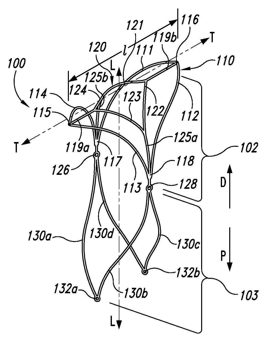
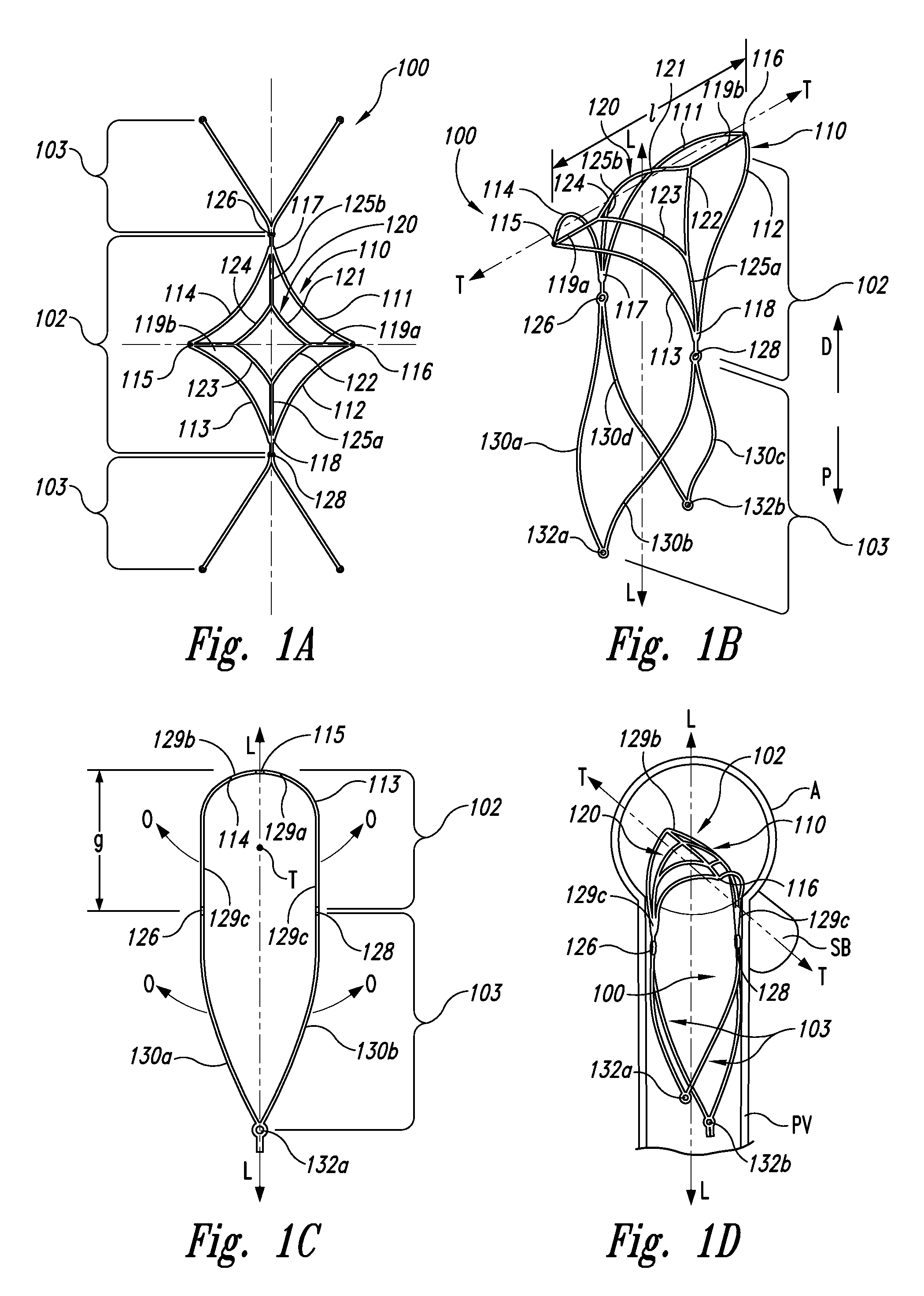
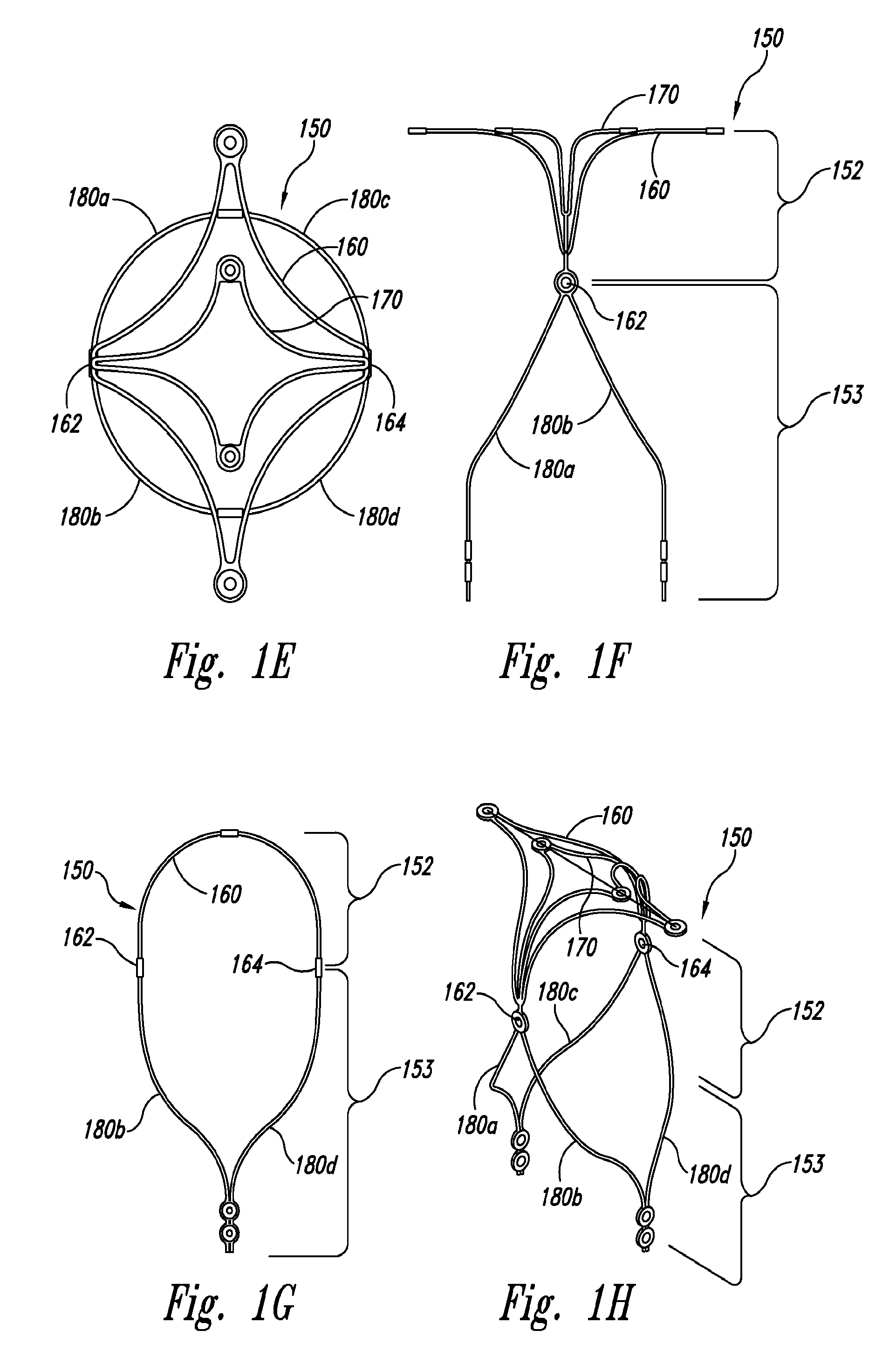
[0041]The presently described technology provides an aneurysm closure device comprising a closure structure and a supplemental stabilizer. The closure structure can have a curved portion configured to extend along a first vessel, such as a side branch of a bifurcated vessel that extends along a lateral axis. The curved portion can have an arch with a proximal-facing surface curved about the lateral axis along the first vessel and a distal-facing surface configured to extend across at least a portion of a neck of an aneurysm at the first vessel. The curved portion of the closure structure can be further configured to exert a radially outward force against the first vessel. The supplemental stabilizer extends from the closure structure along a longitudinal axis transverse to the lateral axis of the first vessel. The supplemental stabilizer is configured to exert an outward force against a second vessel, such as a parent vessel, that extends transversely to the first...


PUM


Abstract

Implantable therapeutic devices and methods for endovascular placement of devices at a target site, such an opening at a neck of an aneurysm, are disclosed. Selected embodiments of the present technology have closures (102) that at least partially occlude the neck of an aneurysm to stabilize embolic or coagulative treatment of the aneurysm. In one embodiment, for example, an aneurysm closure device comprises a closure structure (102) and a supplemental stabilizer (103). The closure structure can have a curved portion configured to extend along a first vessel, such as a side branch of a bifurcated vessel that extends along a lateral axis (T). The supplemental stabilizer extends from the closure structure along a longitudinal axis (L) transverse to the lateral axis of the first vessel. The supplemental stabilizer is configured to exert an outward force against a second vessel, such as a parent vessel, that extends transversely to the first vessel.

Description

CROSS-REFERENCE TO RELATED APPLICATIONS[0001]The present application claims the benefit of the following pending applications:[0002](a) This application is a U.S. National Stage of International Application No. PCT / US10 / 47908, filed Sep. 3, 2010, entitled “SYSTEMS AND METHODS FOR ENCLOSING AN ANATOMICAL OPENING”;[0003](b) U.S. Provisional Patent App. No. 61 / 240,180, filed on Sep. 4, 2009; and[0004](c) U.S. Provisional Patent App. No. 61 / 313,096, filed on Mar. 11, 2010.[0005]All publications and patent applications mentioned in this specification are herein incorporated by reference in their entireties to the same extent as if each individual publication or patent application was specifically and individually indicated to be incorporated by reference.TECHNICAL FIELD[0006]The present technology relates to implantable therapeutic devices and methods for endovascular placement of devices at a target site, such an opening at a neck of an aneurysm. Selected embodiments of the present tech...

Claims


Application Information

Patent Timeline
Patent Type & Authority Patents(United States)
IPC IPC(8): A61B17/12A61B17/00
CPCA61B17/12113A61B17/1214A61B17/1219A61B17/12022A61B17/12118A61B17/12172A61B2017/00632A61B2017/00867A61B2017/12054A61B17/12031A61B17/1215A61B2017/12004A61F2/82A61F2/966A61B17/12145
Inventor CLARKE, GILBERTGERBERDING, BRENTABRAMS, ROBERT M.MOLAEI, MASOUD
Owner PULSAR VASCULAR
Why Eureka
Try Eureka