Geographic atrophy identification and measurement

a geographic atrophy and identification technology, applied in the field of identification and measurement of geographic atrophy, can solve the problems of reducing reflectivity, thinning affected tissues, and increasing thickness

Active Publication Date: 2016-12-27
KK TOPCON
View PDF30 Cites 20 Cited by
  • Summary
  • Abstract
  • Description
  • Claims
  • Application Information

AI Technical Summary

Problems solved by technology

Changes in thickness tend to result in thinning of the affected tissues with disease progression; however, less common retinal diseases related to inflammation can cause an increase in thickness.
Alternatively and spuriously, decreases in reflectivity can result from increased shadowing (darkening, reducing the dynamic range of data) associated with lens crystallizations (cataracts) or floaters in the vitreous.
Retinitis pigmentosa is a progressive retinal disease that affects the photoreceptors resulting in a severe loss of vision.
This is also related to failure to clear some metabolites from outer segment phagocytosis from the RPE.
However, there are a number of problems with current techniques for quantifying GA.
For the sub-RPE slab technique, the complexity of the OCT signal in the choroid can lead to some degree of randomness in the integrated signal and resulting analysis.
These deficiencies can produce complexities that make it difficult for image processing algorithms to effectively and robustly detect atrophic regions.
In addition to the above, ANSI, and possibly other standards organizations as well, places safety limits on the maximum power density that can be applied to the ocular surfaces such as the cornea and retina.
This means that there is an effective limit with respect to the achievable sensitivity for in vivo ocular imaging.
With this in mind, it is not possible to arbitrarily increase the light power in order to achieve any desired signal intensity at deeper retinal positions.

Method used


Image

  • Geographic atrophy identification and measurement
  • Geographic atrophy identification and measurement
  • Geographic atrophy identification and measurement

Examples


Embodiment Construction

[0040]Certain terminology is used herein for convenience only and is not to be taken as a limitation on the present invention. Relative language used herein is best understood with reference to the drawings, in which like numerals are used to identify like or similar items. Further, in the drawings, certain features may be shown in somewhat schematic form.

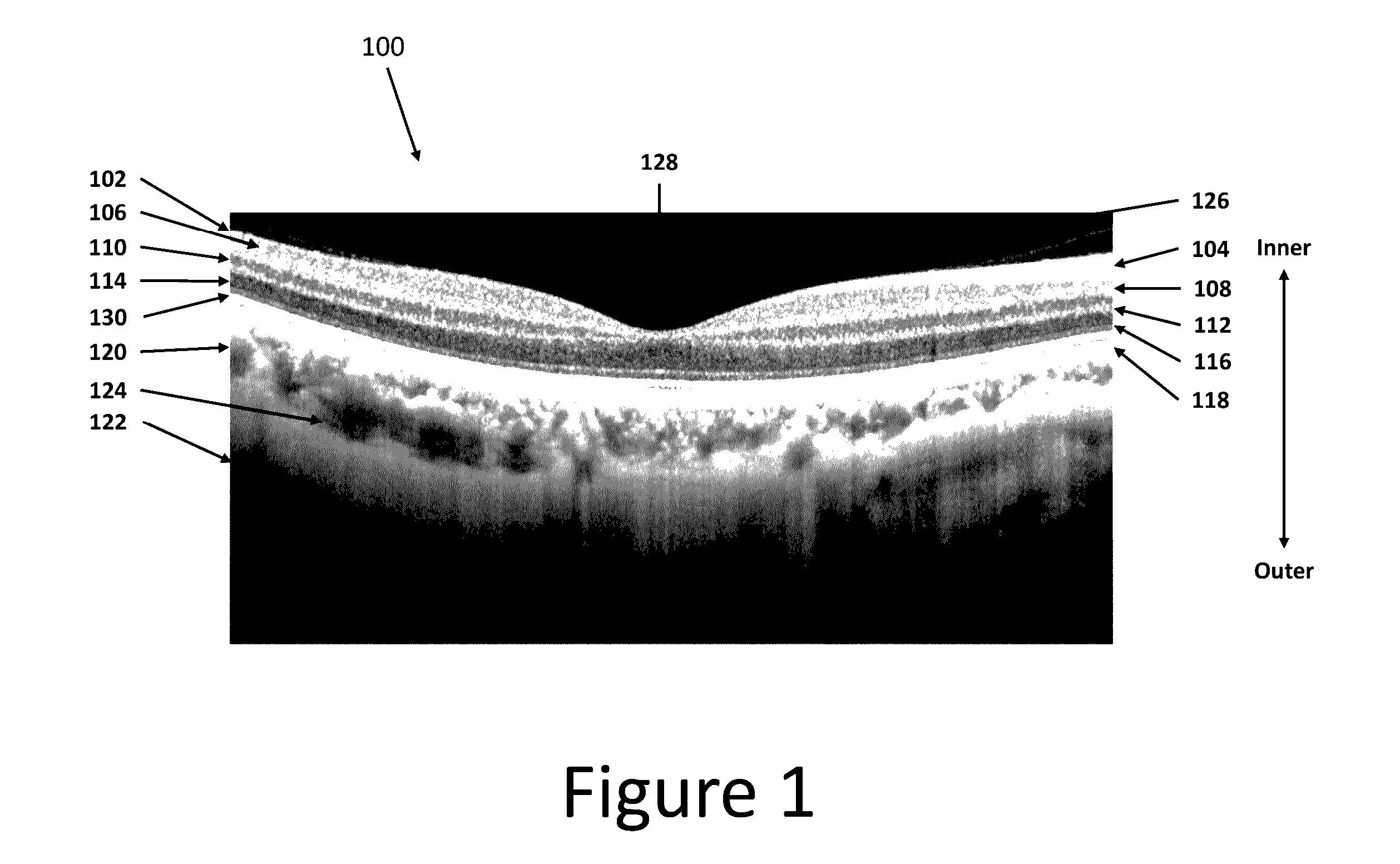
[0041]It should be noted that while this disclosure is based on the OCT imaging modality, it is not limited to OCT. For example, it is applicable to ultrasound as well. It should also be noted that the term “retina” typically refers to the tissue inner to the retinal pigment epithelium (RPE) cell bodies. For the purposes of this disclosure, the retina is defined as the tissue layers including and inner to the RPE cell bodies. Therefore, the choroid and sclera are not considered retinal tissue as used herein. A more detailed illustration of the anatomy and relative locations of layers of the eye and retina as referred to herein is s...


PUM

PropertyMeasurementUnit
center wavelengthaaaaaaaaaa
wavelengthaaaaaaaaaa
wavelengthsaaaaaaaaaa

Abstract

Geographic atrophy of the eye can be detected and measured by imaging the eye at a depth greater than the retinal pigment epithelium (RPE) at a plurality of locations of the eye, for example, using optical coherence tomography (OCT); determining a ratio of the intensities of imaging signals of a retinal layer(s) with respect to the intensity of imaging signals of a sub-RPE layer(s) at each location; determining representative values based at least in part on the determined ratios; generating a map of the representative values; and identifying diseased areas from the map. Contours and binary maps may be generated based on the identified diseased areas. The size and shape of the identified areas may be analyzed and monitored over a period of time.

Description

CROSS REFERENCE TO RELATED APPLICATIONS[0001]This application claims priority to U.S. Provisional Application Ser. No. 61 / 929,743, filed on Jan. 21, 2014, entitled “GEOGRAPHIC ATROPHY IDENTIFICATION AND MEASUREMENT”, the entirety of which is incorporated herein by reference.BACKGROUND OF THE INVENTION[0002]1. Field of the Invention[0003]This application relates generally to the identification and measurement of geographic atrophy, and more specifically, to the identification of geographic atrophy in ophthalmic optical coherence tomography applications.[0004]2. Description of Related Art[0005]In ophthalmic and other applications, optical coherence tomography (OCT) is frequently used to generate three-dimensional scan data of a volume. OCT scans typically utilize one of relatively few fixation positions—most commonly the macula or optic disc, which place the respective feature at or near the center of the OCT scan. Wide scan patterns, which cover a large area that encompasses both the...

Claims


Application Information

Patent Timeline
Patent Type & Authority Patents(United States)
IPC IPC(8): A61B3/00A61B3/10A61B3/12G01N21/47G06T5/00G01N21/64G06T7/00G01N21/17G01N21/88
CPCA61B3/0025A61B3/102A61B3/12G01N21/4795G01N21/6456G06T5/002G06T7/0012G01N2021/1787G01N2021/887G06T2207/10101G06T2207/30041G06V40/197A61B3/1025A61B3/1225A61B5/4842G01B9/02091
Inventor YANG, QIREISMAN, CHARLES A.
Owner KK TOPCON
Why Eureka
Try Eureka