Internal device, external device, diagnosis apparatus including the same, and endoscope apparatus

a technology of endoscope and capsule, which is applied in the field of internal devices, external devices, and diagnosis apparatus comprising the same, and the same, which can solve the problems of difficult to recognize an accurate location inside the body the need to reduce the resolution of the camera to generate, and the difficulty of the capsule endoscope to recognize an accurate location

Active Publication Date: 2017-07-18
IND ACADEMIC CORP FOUND YONSEI UNIV
View PDF12 Cites 0 Cited by
  • Summary
  • Abstract
  • Description
  • Claims
  • Application Information

AI Technical Summary

Benefits of technology

[0006]The present invention provides an internal device, external device, diagnosis apparatus including the same, and endoscope apparatus that accurately detect the location of an internal device that is inserted into an inside of a body and moves therein, such as a capsule endoscope.
[0007]The present invention provides an internal device, external device, diagnosis apparatus including the same, and endoscope apparatus that control an internal device based on the location of the internal device to enhance control performance.
[0008]The present invention provides an internal device, external device, diagnosis apparatus including the same, and endoscope apparatus that provide information on an internal device to a user such as a patient or medical team based on the location of the internal device to enhance user convenience.
[0015]In further embodiments, the control unit may switch the internal device to an idle mode to decrease power consumption when a power-supply cutoff signal is received from the external device.
[0040]In even further embodiments, the second control unit may generate a power-supply cutoff signal to transmit the signal to the internal device when the region in which the internal device is located corresponds to a first preset target region, and the first control unit may switch the internal device to an idle mode to decrease power consumption when the power-supply cutoff signal is received from the external device.

Problems solved by technology

However, it is difficult for the capsule endoscopes to recognize an accurate location inside a body and the capsule endoscopes continuously capture images of the inside of the body in order to track the location.
However, due to the limited power of the capsule endoscopes, there is a need to decrease the resolution of a camera to generate an image having a low resolution in order to continuously capture images for a long time for which the capsule endoscopes stay at the inside of the body.
As a result, there was typically a limitation in discovering a lesion or diagnosing disease with the capsule endoscopes, and even if the lesion was discovered, there was a limitation in that it was difficult to recognize an accurate location.

Method used


Image

  • Internal device, external device, diagnosis apparatus including the same, and endoscope apparatus
  • Internal device, external device, diagnosis apparatus including the same, and endoscope apparatus
  • Internal device, external device, diagnosis apparatus including the same, and endoscope apparatus

Examples


Embodiment Construction

[0056]Embodiments of the present invention are described below in detail with reference to the drawings attached to the present disclosure.

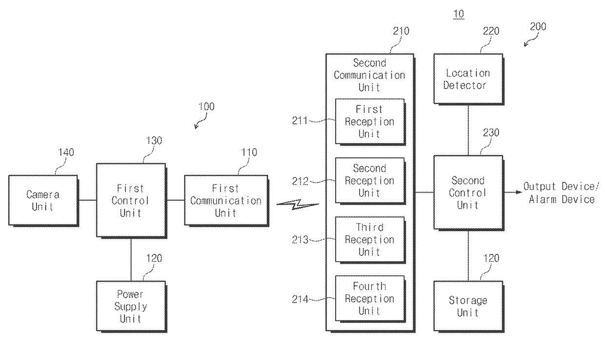
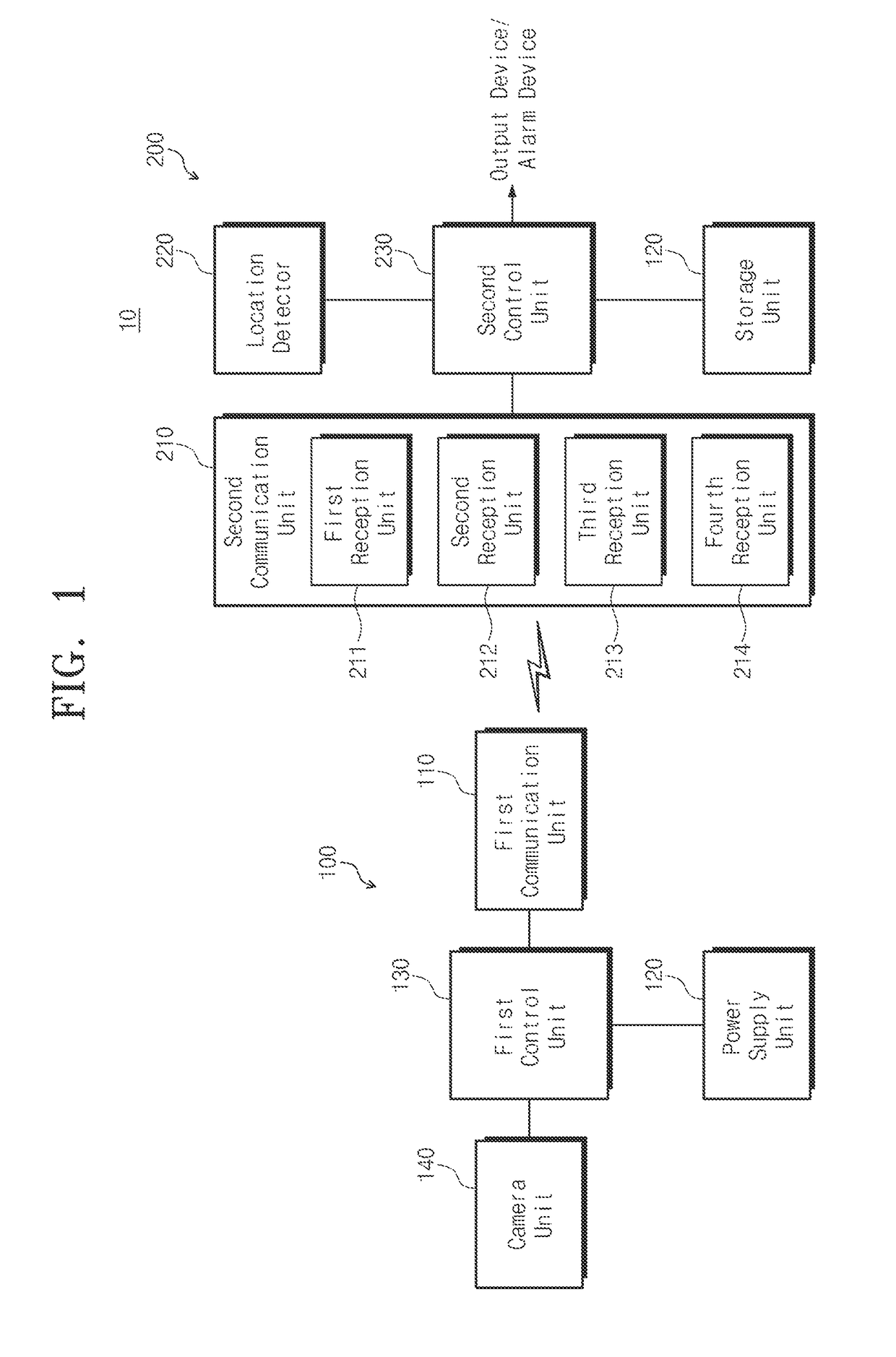
[0057]FIG. 1 is an exemplary block diagram of a diagnosis apparatus 10 according to an embodiment of the present invention.

[0058]As shown in FIG. 1, the diagnosis apparatus 10 may include an internal device 100 and an external device 200. The internal device 100 is inserted into the internal body and moves therein. The external device 200 is located at an outside of the body and controls the internal device 100.

[0059]According to an embodiment, the internal device 100 may be a capsule endoscope that includes a camera to capture images of the internal body. However, the internal device 100 is not limited to the capsule endoscope and includes all apparatuses inserted into the body to be used for diagnosis and therapy, such as a capsule-type surgical instrument including a surgical instrument, or a drug injection instrument injecting drug into an in...


PUM


Abstract

Provided is a diagnosis apparatus. The diagnosis apparatus includes: an internal device inserted into an inside of a body and moving therein; and an external device located at an outside of the body and controlling the internal device, wherein the internal device includes: a first communication unit communicating with the external device; a power supply unit supplying power for driving of the internal device; and a first control unit controlling an operation of the internal device according to a control signal when the control signal is received from the external device, and the external device includes: a second communication unit communicating with the internal device; a location detection unit processing a signal received from the internal device to detect a location of the internal device; and a second control unit generating a control signal for controlling the internal device based on the location of the internal device.

Description

CROSS-REFERENCE TO RELATED APPLICATIONS[0001]This U.S. non-provisional patent application claims priority under 35 U.S.C. §119 of Korean Patent Application No. 10-2014-0011011, filed on Jan. 29, 2014, the entire contents of which are hereby incorporated by reference.BACKGROUND OF THE INVENTION[0002]The present invention disclosed herein relates to an internal device, an external device, a diagnosis apparatus comprising the same, and an endoscope apparatus.[0003]Endoscopes that may immediately observe the inside of an organ or body cavity without an operation or autopsy are divided into general endoscopes that insert tubes having cameras at their ends into insides of bodies, and capsule endoscopes that patients swallow like tablets so that the endoscopes are inserted into the insides of the bodies. While general endoscopes causes pain and displeasure to patients when examination is performed, the capsule endoscopes supplement such a disadvantage of the general endoscopes and have an ...

Claims


Application Information

Patent Timeline
Patent Type & Authority Patents(United States)
IPC IPC(8): A61B1/04A61B1/00A61B5/06H04N5/225
CPCA61B1/00006A61B1/00036A61B1/041A61B5/061H04N2005/2255H04N23/555
Inventor KIM, TAE WOOK
Owner IND ACADEMIC CORP FOUND YONSEI UNIV
Why Eureka
Try Eureka