Methods and compositions for isolating exosomes

a technology of exosomes and compositions, applied in the field of biotechnology, can solve the problems of time-consuming, cumbersome and/or costly, limited by the amount of material that can be processed, and achieve the effects of rapid, efficient and cost-effectiv

Inactive Publication Date: 2017-12-05
BOARD OF RGT THE UNIV OF TEXAS SYST +1
View PDF8 Cites 6 Cited by
  • Summary
  • Abstract
  • Description
  • Claims
  • Application Information

AI Technical Summary

Benefits of technology

[0012]The present invention addresses the foregoing and other needs of the prior art by providing new methods and compositions for isolating extracellular microvesicles such as exosomes, particularly disease-related and phosphatidylserine (PS)-positive exosomes, including tumor-derived exosomes, which methods are rapid, efficient, cost-effective and suitable for use with large volumes of biological fluids. These methods are based on the surprising use of acetate buffers to isolate extracellular microvesicles such as exosomes from solution. The invention provides the ability to isolate large quantities of extracellular microvesicles and exosomes, particularly antigenically intact, disease-related and PS-positive exosomes such as tumor-derived exosomes, which are indistinguishable from those prepared by the current ultracentrifugation methods, the latter of which are time-consuming, cumbersome and volume-limited.
[0019]By applying the invention to all disease-related extracellular microvesicles, such as disease-related exosomes, phosphatidylserine will preferably be present on the surface of the extracellular microvesicles, more preferably in association with non-lipid membrane components, wherein the non-lipid membrane components comprise membrane proteins. In this regard, the acetate buffer is believed to neutralize the surface charge of the phosphatidylserine on the disease-related extracellular microvesicles, thereby precipitating the disease-related extracellular microvesicles from the biological fluid.
[0034]As the invention effectively separates disease-related extracellular microvesicles from biological fluids that contain mixed populations of extracellular microvesicles, the invention further provides methods to obtain disease-related, preferably tumor-derived, extracellular microvesicles from the supernatant of diseased or tumor cells cultured in the presence of serum that contains normal extracellular microvesicles, preferably wherein the disease-related or tumor-derived extracellular microvesicles have negatively-charged phosphatidylserine on their surface. These methods comprise contacting the supernatant with an acetate buffer at a pH and concentration effective to selectively precipitate disease-related, preferably tumor-derived, extracellular microvesicles from the supernatant and collecting the precipitate, thereby obtaining disease-related, preferably tumor-derived, extracellular microvesicles from the supernatant without substantial contamination from normal extracellular microvesicles in the serum. A particular example is wherein mouse or human tumor cells are cultured in the presence of bovine or fetal bovine serum.
[0059]Acetate buffers comprising both sodium acetate and potassium acetate, and mixtures thereof, are effective. In certain embodiments, the acetate buffers are essentially free from volume excluding polymers, such as polyethylene glycol. An exemplary preferred embodiment is a sodium acetate buffer that has a pH of about 4.75 and a final concentration in the sample of about 0.1M.

Problems solved by technology

However, the available techniques are limited by drawbacks in two important respects.
Firstly, as applied to extracellular microvesicle and exosome preparation in general, they are time-consuming, cumbersome and / or costly, and limited by the amounts of material that can be processed.
The typical approach of concentrating the biological medium using ultracentrifugation before proceeding with exosome isolation is very time consuming and requires specialized laboratory equipment.
Secondly, the current techniques are particularly limited as they apply to tumor-derived extracellular microvesicle and exosomes.
In addition to the general drawbacks described above, such methods and kits are very limiting, requiring both advance knowledge of a particular exosome surface marker to be exploited in the antibody binding, as well as many detailed technical steps in the protocol, such as the preparation and use of biotinylated capture antibodies.

Method used


Image

  • Methods and compositions for isolating exosomes
  • Methods and compositions for isolating exosomes
  • Methods and compositions for isolating exosomes

Examples


example i

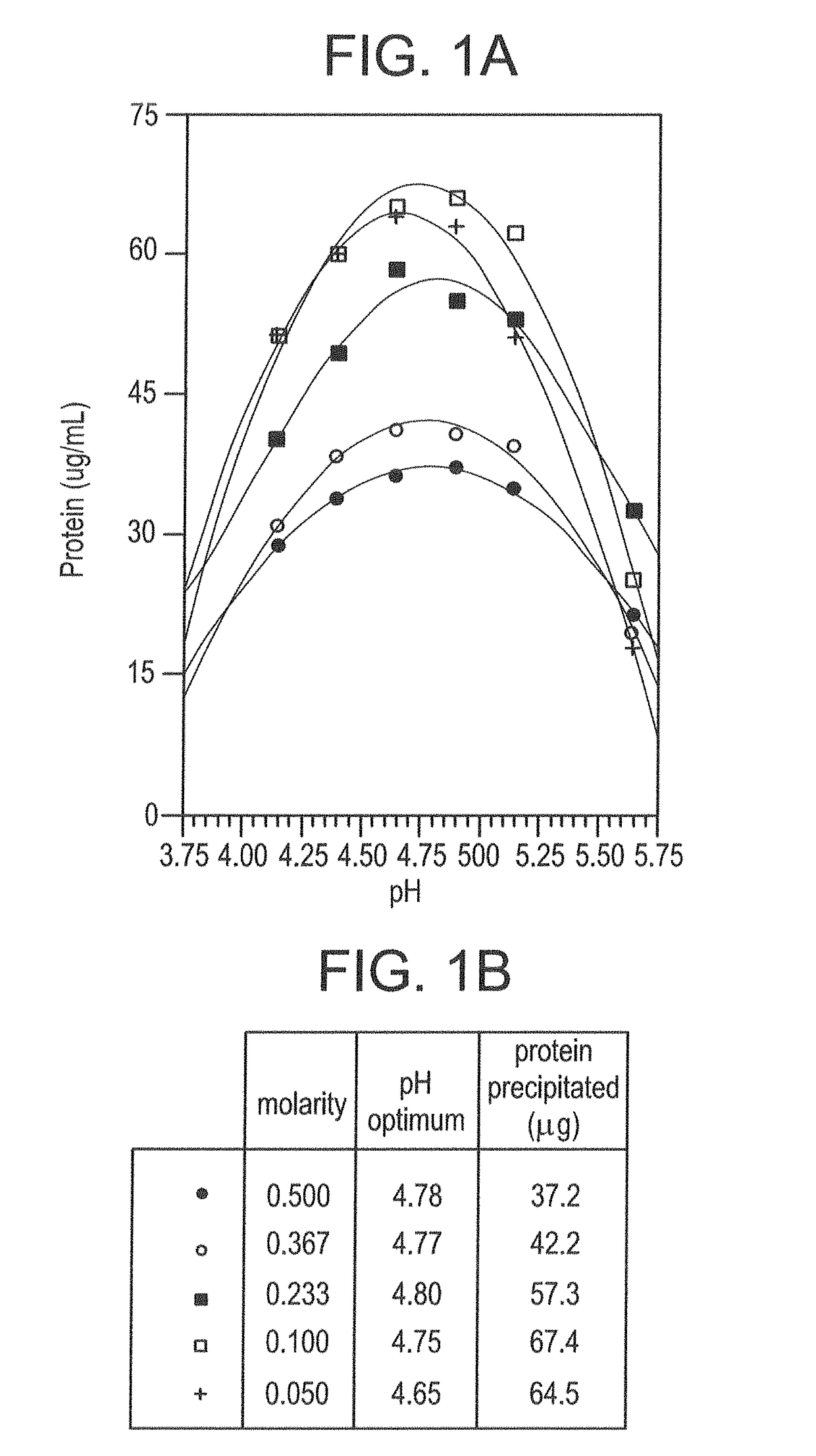
Isolating Tumor-Derived Exosomes Using Acetate Buffers

[0149]The present example shows advantageous methods for isolating extracellular microvesicles such as exosomes, particularly tumor-derived exosomes, using acetate buffers.

A. Materials and Methods

[0150]The following Materials and Methods are relevant to the results reported in Example I, Example II and Example III.

[0151]1. Tissue Culture

[0152]K1735P murine melanoma (tumor) cells (provided by I. J. Fidler, M. D. Anderson Cancer Center, Houston, Tex., but anyway widely available) were cultured in minimal essential media (MEM) supplemented with L-glutamine (2 mM), Na pyruvate (1 mM), penicillin (100 U / mL), streptomycin (100 μg / mL), nonessential amino acids and fetal bovine serum (10%). Cells (about 25×106 in 15 mL media) were seeded into the lower chamber of CELLine AD 1000 flasks (Integra Biosciences AG) that contained 250 mL media in the upper chamber (Mitchell et al., 2008). Conditioned media (about 15 mL) was collected from the ...

example ii

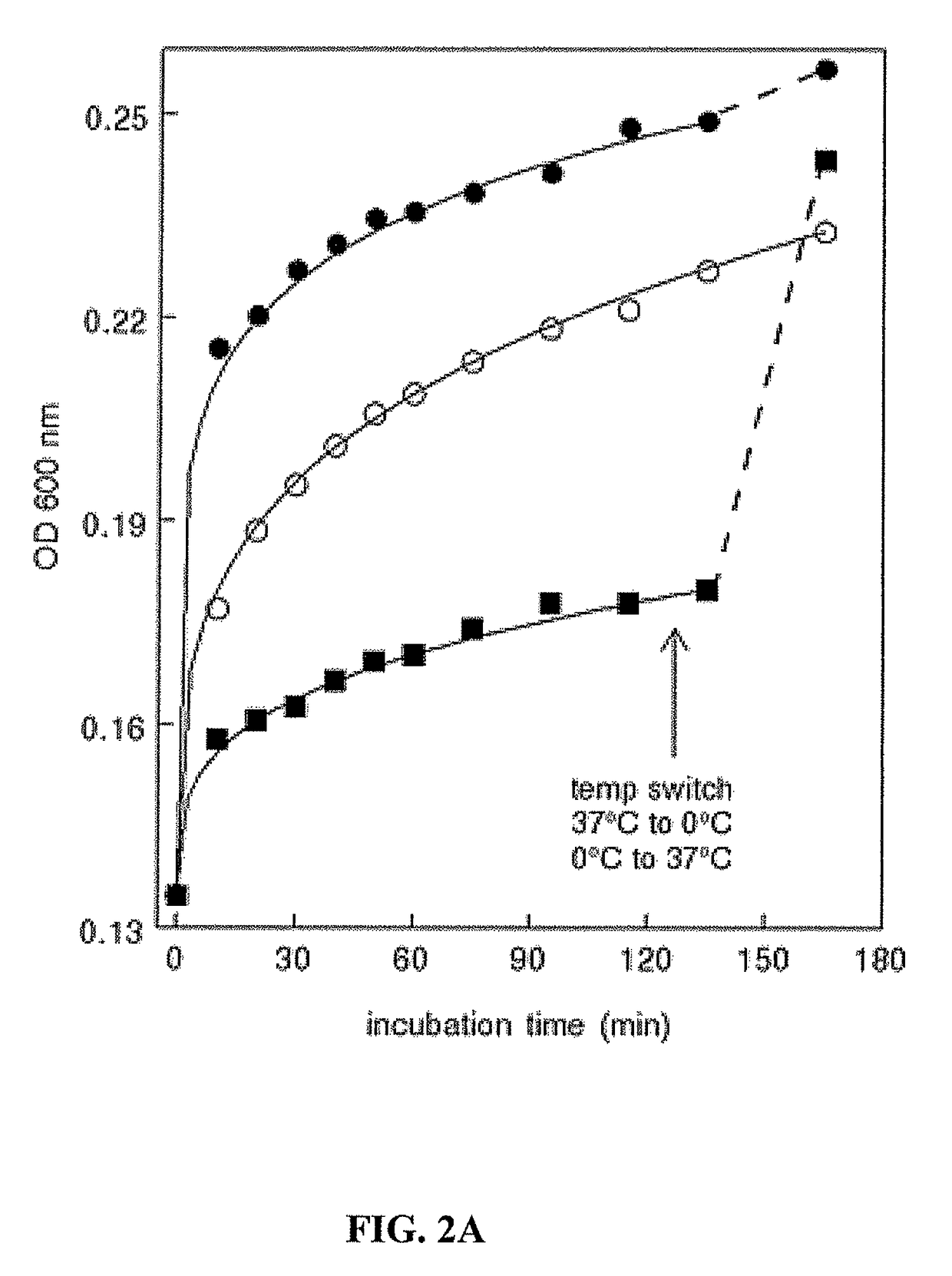
Further Characterization of Tumor Exosome Isolation

[0169]This example further characterizes the isolation methodology for disease-related extracellular microvesicles such as exosomes, and highlights the importance of acetate buffers in the technique with reference to tumor exosomes.

A. Temperature Independence

[0170]The present study shows that tumor exosome precipitation is essentially temperature-independent. However, analyzing the effect of temperature showed that the development of turbidity was temperature-dependent.

[0171]An immediate temperature-dependent increase in turbidity occurred upon the addition of acetate, which then began to level off. Continued incubation showed a modest, about 2-fold increase in rate between 0° C. and 20° C. However, no significant difference in rate was observed upon increasing the temperature from 20° C. to 37° C. (FIG. 2A). Interestingly, once the reaction plateaued at 0° C., increasing the temperature to 37° C. resulted in an immediate increase i...

example iii

Equivalent Yield of Morphologically Verified Tumor Exosomes

[0179]The present example shows that the yield of disease-related extracellular microvesicles such as exosomes isolated using acetate buffers is equivalent to that from the typical ultracentrifugation method, even though the acetate method is easier and quicker. The acetate-purified exosomes, as exemplified by tumor exosomes, are also shown to be morphologically indistinguishable from those prepared by traditional ultracentrifugation and to be antigenically intact.

A. Equivalent Yield

[0180]The relative yield of tumor exosomes obtained with acetate isolation and with conventional 100,000 g ultracentrifugation was quantified by assessing alix and PS in both populations. Flow cytometry analysis of alix with Cy3-labeled alix antibodies (FIG. 5A) and PS with FITC-labeled annexin 5 (FIG. 5B) suggested that both methods yielded similar amounts of exosomes.

[0181]In directly comparing the yield of protein obtained with acetate isolati...


PUM

PropertyMeasurementUnit
volumeaaaaaaaaaa
pHaaaaaaaaaa
temperatureaaaaaaaaaa

Abstract

Disclosed are surprising new methods and compositions for isolating extracellular microvesicles such as exosomes, particularly disease-related and phosphatidylserine (PS)-positive extracellular microvesicles as exemplified by tumor- and viral-derived exosomes. The methods of the invention are rapid, efficient, cost-effective and, importantly, are suitable for use with large volumes of biological fluids and produce antigenically intact extracellular microvesicles and exosomes. The methods and compositions are based on the surprising use of acetate buffers to isolate large quantities of extracellular microvesicles, particularly tumor-derived exosomes, from solution, without damaging their morphological or functional properties or antigenicity.

Description

CROSS-REFERENCE TO RELATED APPLICATIONS[0001]The present application claims priority to first U.S. provisional application Ser. No. 61 / 945,718, filed Feb. 27, 2014, and to second U.S. provisional application Ser. No. 61 / 970,529, filed Mar. 26, 2014, the entire disclosures of which applications, including the specification, claims and drawings are incorporated herein by reference without disclaimer.BACKGROUND OF THE INVENTION[0002]Field of the Invention[0003]The present invention relates to the fields of biotechnology, and particularly to extracellular microvesicles such as exosomes. The invention provides surprising new methods and compositions for isolating disease-related and phosphatidylserine (PS)-positive extracellular microvesicles such as tumor- and viral-derived exosomes, particularly tumor-derived exosomes, which are especially suitable for use with large volumes of biological fluids and produce extracellular microvesicles and exosomes that are antigenically intact.[0004]De...

Claims


Application Information

Patent Timeline
Patent Type & Authority Patents(United States)
IPC IPC(8): G01N33/53G01N33/574G01N33/50
CPCG01N33/574G01N33/57484G01N33/5076
Inventor SCHROIT, ALAN J.THORPE, PHILIP E.FUSSEY, SHELLEY P. M.
Owner BOARD OF RGT THE UNIV OF TEXAS SYST
Why Eureka
Try Eureka