Semi-automatic partition method of lung CT image focus

A CT image, semi-automatic technology, applied in image enhancement, image data processing, instruments, etc., can solve the problems of low efficiency and poor accuracy of manual lesion segmentation, and achieve the effect of good practicability, accurate segmentation, and high efficiency

Inactive Publication Date: 2009-12-23
XIAN UNIV OF TECH
View PDF5 Cites 2 Cited by
  • Summary
  • Abstract
  • Description
  • Claims
  • Application Information

AI Technical Summary

Problems solved by technology

[0003] The purpose of the present invention is to provide a semi-automatic segmentation method for lung CT image lesions, which solves the shortcomings of low efficiency and poor accuracy of manual focus segmentation

Method used


Image

  • Semi-automatic partition method of lung CT image focus
  • Semi-automatic partition method of lung CT image focus
  • Semi-automatic partition method of lung CT image focus

Examples


Embodiment Construction

[0017] The present invention will be described in detail below by means of drawings and examples.

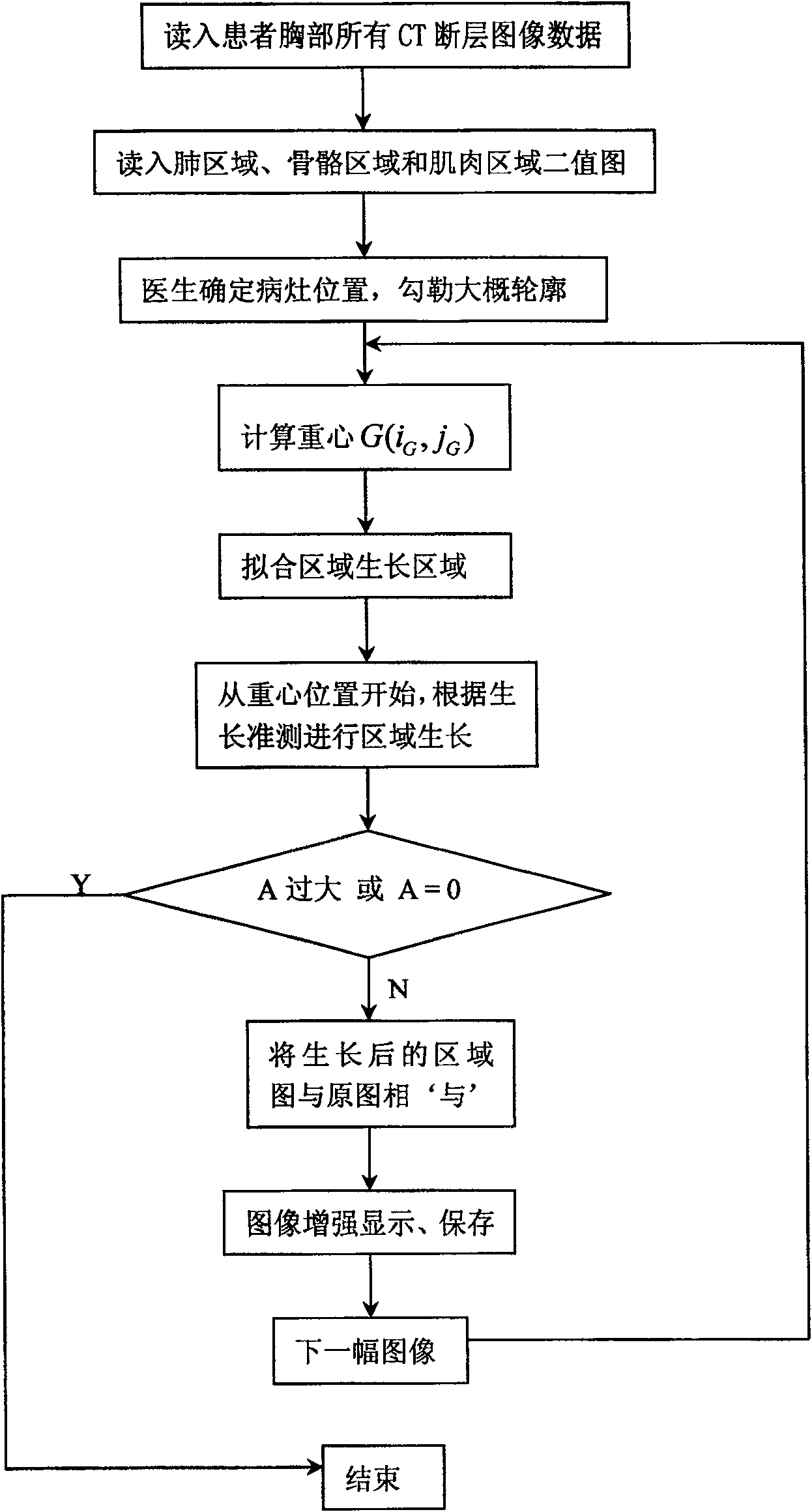
[0018] The method of the present invention presses figure 1 The flow shown is carried out, and the following is an embodiment provided by the method of the present invention.

[0019] First, the CT equipment reads in all the CT tomographic image data of the patient's chest, and then proceeds to the following steps,

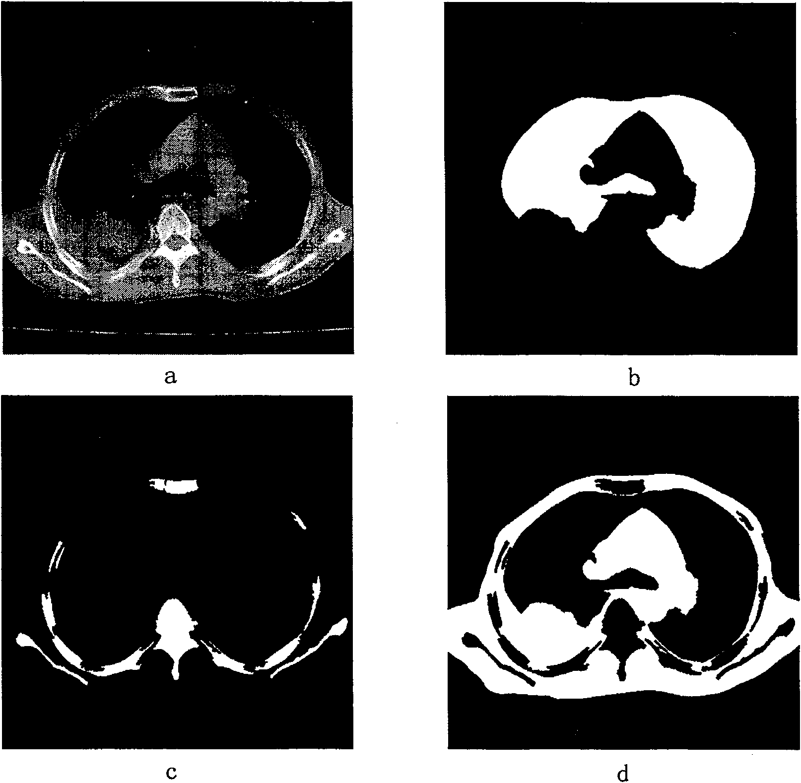
[0020] a) Segment lung region, bone region and muscle region

[0021] In all the CT tomographic image data of the chest, according to the principle of CT imaging and the different CT values ​​of each tissue, the lung area, bone area and muscle area are segmented, that is, the lung area with a CT value distribution range of -900Hu to -100Hu is segmented out, segment the muscle area with a CT value distribution range of -30Hu to 100Hu, and segment the bone area with a CT value distribution range of 100Hu or more, and obtain their binary images respectively, as shown...


PUM


Abstract

The method for semi-automatic segmentation of lung CT image lesions disclosed in the present invention firstly divides the lung area, bone area and muscle area according to the data of all CT tomographic images of the patient's chest read by CT equipment, and initially determines the approximate location and range of the lesion to limit The region growing method segmented the lesion area of ​​other slices, and then enhanced the image of the segmented lesion area. The method of the present invention extracts data from each tomographic lesion based on the inherent characteristics that the same tissue has similar CT values ​​and the lesion body has continuity in three-dimensional space. The method is accurate and efficient for lesion segmentation, and has good practicability .

Description

technical field [0001] The invention belongs to the technical field of automatic processing of CT images, and relates to an image segmentation technology, in particular to a semi-automatic segmentation method for lung CT image lesions. Background technique [0002] The lesion segmentation and enhancement technology of CT images currently mainly relies on doctors to manually outline the lesions on all 2D tomographic images containing lesions. Doctors need to spend a lot of time on repetitive manual lesion outlining work, which is inefficient, and the repeated outlining work is easy to make doctors tired, and misoperations are prone to occur when tired. Contents of the invention [0003] The purpose of the present invention is to provide a semi-automatic segmentation method for lung CT image lesions, which solves the shortcomings of low efficiency and poor accuracy of manual lesion segmentation. [0004] The technical scheme adopted in the present invention is that the lung...

Claims


Application Information

Patent Timeline
Patent Type & Authority Patents(China)
IPC IPC(8): G06T5/00A61B6/03
Inventor 朱虹邓杰航马湘旺张波
Owner XIAN UNIV OF TECH
Why Eureka
Try Eureka