Recognition and counting method for cells

A counting method and cell technology, applied in the field of medical image processing, can solve problems such as low efficiency, doping with subjective factors, and unsatisfactory cell identification methods

Active Publication Date: 2016-10-26
HEFEI UNIV OF TECH
View PDF5 Cites 35 Cited by
  • Summary
  • Abstract
  • Description
  • Claims
  • Application Information

AI Technical Summary

Problems solved by technology

This method has many deficiencies: firstly, the inspection workload is large, the efficiency is low, and continuous work is easy to cause wrong identification due to objective factors; secondly, the identification and analysis of samples is easily restricted by visual fatigue, etc....

Method used


Image

  • Recognition and counting method for cells
  • Recognition and counting method for cells
  • Recognition and counting method for cells

Examples


Embodiment

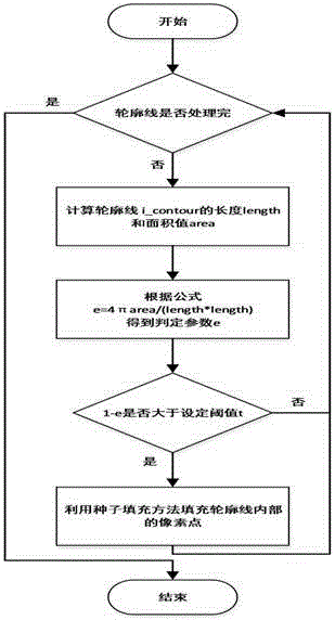
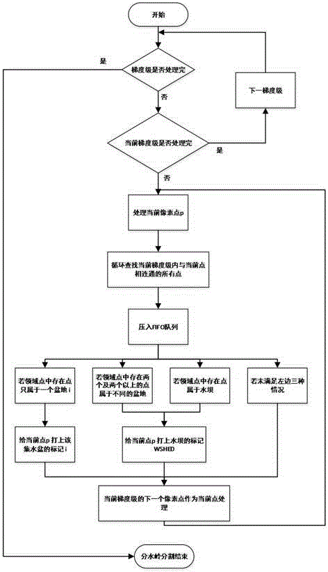
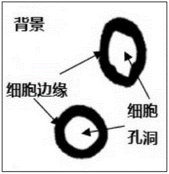
[0135] More specifically, as figure 1 , figure 2 and image 3 Shown, the present invention comprises the following steps:

[0136] 1. Preprocessing stage of image acquisition

[0137] The image that is collected is carried out gray scale, uses weighted average method, three components are carried out weighted average with different weights, obtains gray scale image, the weighting formula used in the present invention is as follows:

[0138] f(i,j)=0.30R(i,j)+0.59G(i,j)+0.11B(i,j)

[0139] f(i,j) represents the grayscale value of the image after grayscale, R(i,j), G(i,j), B(i,j) represent the pixel points of the original image before grayscale The grayscale value of the color for the three channels.

[0140] Divide the grayscaled image into 4 equal parts, a total of four rectangular ROI areas, namely img1 in the upper left corner, img2 in the upper right corner, img3 in the lower left corner, and img4 in the lower right corner. The specific parameters are as follows:

...


PUM


Abstract

The invention discloses a recognition and counting method for cells, comprising nine steps: preprocessing an image in a micron-order microscopic acquisition environment; extracting cell holes from the preprocessed image; performing closed hole filling of cells by using the knowledge of a connected domain; extracting a contour point sequence of cells from the image filled in step 3; filling non-closed holes of cells by adopting a non-closed hole filling method based on circularity determination; performing chamfer distance transformation on the filled image; performing extreme value uniqueness marking on cell hole positions; segmenting the image after extreme value uniqueness by using a marked watershed method; and quantifying and marking the segmented result. The method has the advantages that the influence of image noise can be greatly reduced, the phenomena of over-segmentation and discontinuous segment lines are eliminated, the segmentation effect is improved, and the cell recognition rate is improved.

Description

technical field [0001] The invention relates to the technical field of medical image feature extraction and identification in the field of medical image processing, and in particular to a method for identifying and counting cells. Background technique [0002] The methods of the prior art require a large amount of time to examine a sample. This method has many shortcomings: firstly, the inspection workload is large, the efficiency is low, and continuous work is easy to cause wrong identification due to objective factors; secondly, the identification and analysis of samples is easily restricted by visual fatigue, etc., mixed with strong subjective factors without objective criteria. The existing semi-automatic identification and detection methods are becoming more and more inappropriate. When the peak period of detection is encountered, the test results cannot be obtained in time and accurately, which will delay the patient's visit to the doctor. Therefore, realizing the au...

Claims


Application Information

Patent Timeline
IPC IPC(8): G06K9/34G06K9/38G06K9/46
CPCG06V10/267G06V10/28G06V10/457G06V2201/03
Inventor 霍星檀结庆荆珏华董周樑汪国新何逸飞沈宏伟邵堃
Owner HEFEI UNIV OF TECH
Why Eureka
Try Eureka