Ultrasonic diagnosis device, ultrasonic image analysis device, and ultrasonic image analysis method
A diagnostic device and image analysis technology, applied in echo tomography and other directions, which can solve problems such as inability to perform correct settings and skewed myocardial tissue
- Summary
- Abstract
- Description
- Claims
- Application Information
AI Technical Summary
Problems solved by technology
Method used
Image
Examples
no. 1 approach
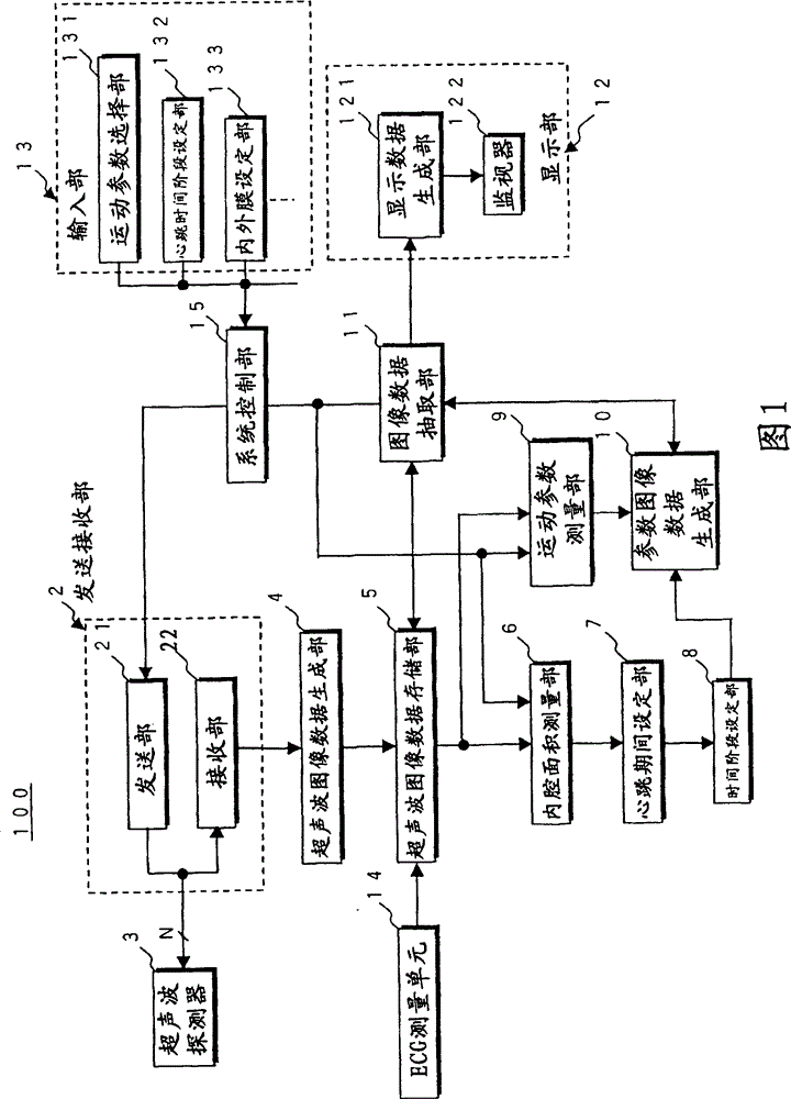
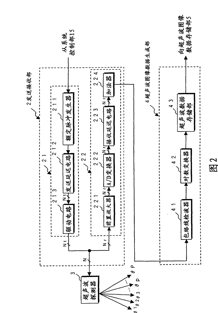
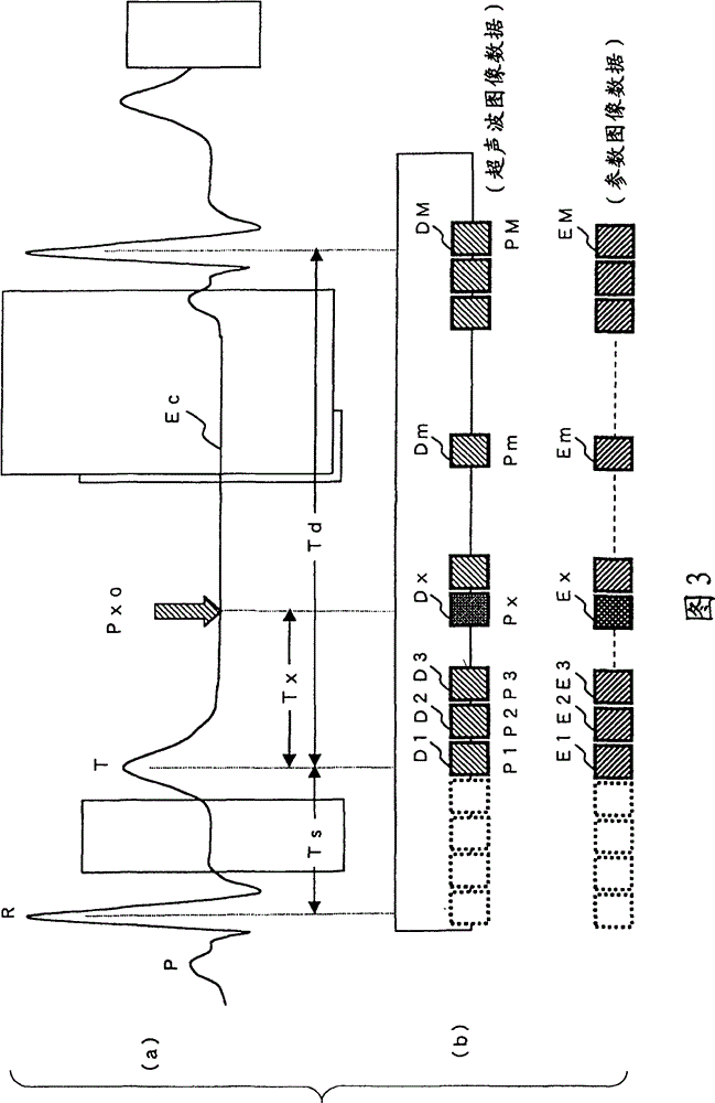
[0023] The ultrasonic diagnostic apparatus in the first embodiment of the present invention described below first transmits and receives ultrasonic waves to the subject to generate time-series B-mode image data as ultrasonic image data, and performs tracking processing on these ultrasonic image data to obtain The "skew (strain)" of myocardial tissue is measured two-dimensionally or three-dimensionally as a motion parameter. On the other hand, the end-systolic phase determined from the time period at which the area of the cardiac chamber becomes the smallest using the ultrasonic image data, and the R wave in the electrocardiographic waveform of the subject measured in parallel with the collection of the ultrasonic image data For the determined end-diastole, set the diastolic heartbeat time phase based on the above-mentioned end-systolic phase, and add the diastolic heartbeat time phase to the time-series parameter image data generated based on the motion parameters of the ultr...
no. 2 approach
[0075] Next, a second embodiment of the present invention will be described. The ultrasonic image analysis device in the second embodiment first performs tracking processing on time-series ultrasonic image data collected in advance with R-wave timing information based on the electrocardiographic waveform of the subject, and two-dimensionally measures Motion parameters of myocardial tissue. On the other hand, the end-systolic phase specified based on the time period at which the area of the cardiac chamber becomes the smallest using the ultrasonic image data and the end-diastolic phase specified using the R-wave timing information added to the ultrasonic image data are set based on the end-systolic phase. The time phase of the diastolic heartbeat is added to the time-series parameter image data generated based on the motion parameters of the ultrasonic image data. Then, the parametric image data of the diastolic heartbeat time phase closest to the desired diastolic heartbeat...
PUM
 Abstract
Description
Claims
Application Information
 - R&D
- Intellectual Property
- Life Sciences
- Materials
- Tech Scout
- Unparalleled Data Quality
- Higher Quality Content
- 60% Fewer Hallucinations
Browse by: Latest US Patents, China's latest patents, Technical Efficacy Thesaurus, Application Domain, Technology Topic, Popular Technical Reports.
© 2025 PatSnap. All rights reserved.Legal|Privacy policy|Modern Slavery Act Transparency Statement|Sitemap|About US| Contact US: help@patsnap.com


