
Ultrasonographic device, ultrasonographic method, and ultrasonographic device data processing program

一种诊断装置、超声波的技术,应用在血流量测量装置等方向,能够解决不自然的图像、无法进行展开、处理复杂运算时间等问题,达到降低噪声的影响、良好且稳定混叠校正的效果

Active Publication Date: 2010-03-24
TOSHIBA MEDICAL SYST CORP
View PDF5 Cites 2 Cited by
  • Summary
  • Abstract
  • Description
  • Claims
  • Application Information

AI Technical Summary

Problems solved by technology

[0015] However, in the conventional method of performing local expansion processing two-dimensionally, there is a problem that calculation time is required due to complicated processing.
In addition, there is a problem that when the number of residues increases, the points that avoid the residues become unnatural images.
Furthermore, there is a problem in robustness, and there is also a problem that expansion cannot be performed sometimes.

Method used


Image

  • Ultrasonographic device, ultrasonographic method, and ultrasonographic device data processing program
  • Ultrasonographic device, ultrasonographic method, and ultrasonographic device data processing program
  • Ultrasonographic device, ultrasonographic method, and ultrasonographic device data processing program

Examples


Embodiment Construction

[0031] Embodiments of the ultrasonic diagnostic apparatus, ultrasonic diagnostic method, and data processing program of the ultrasonic diagnostic apparatus of the present invention will be described with reference to the drawings.

[0032] (structure and function)

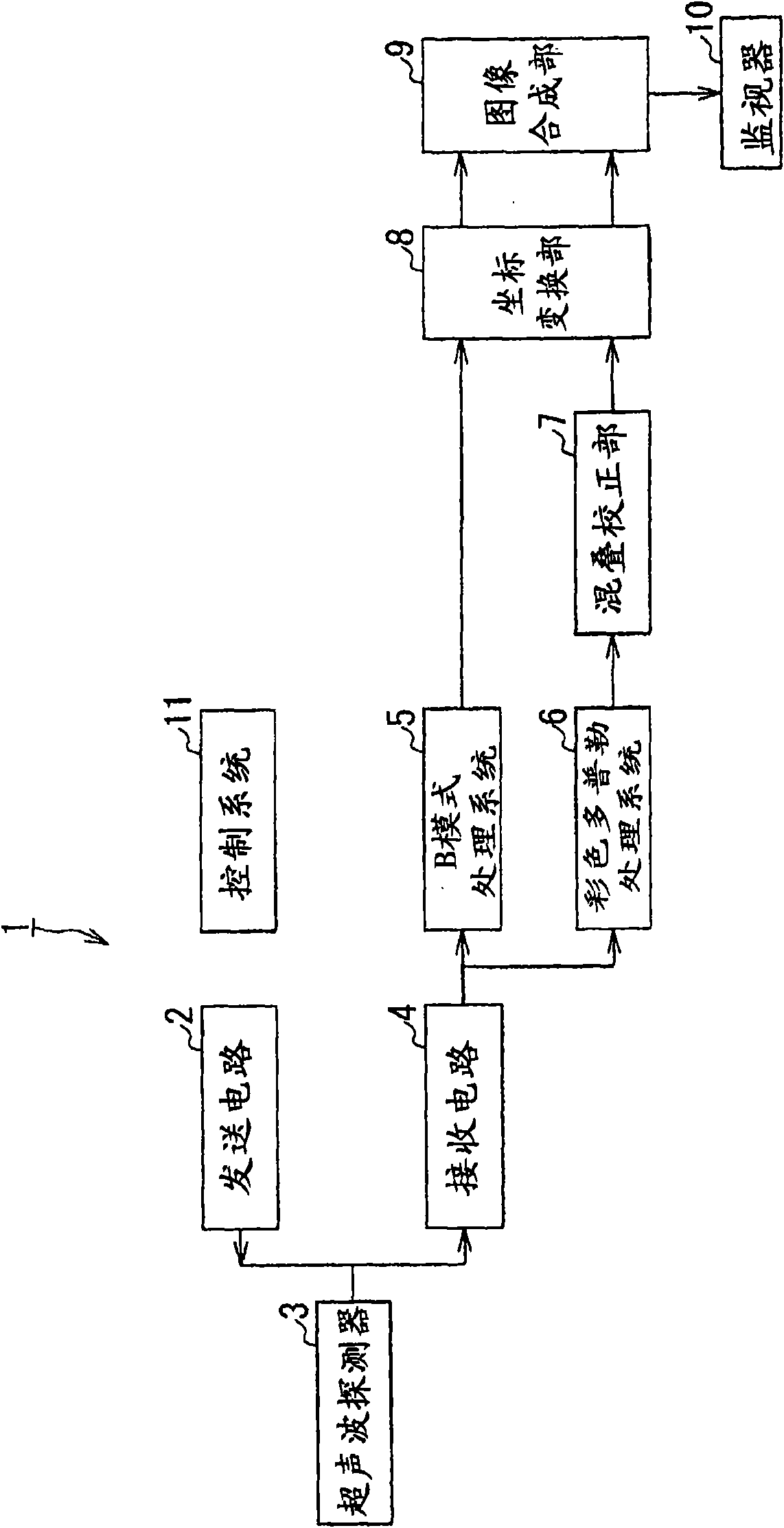
[0033] figure 2 It is a block configuration diagram showing an embodiment of the ultrasonic diagnostic apparatus of the present invention.

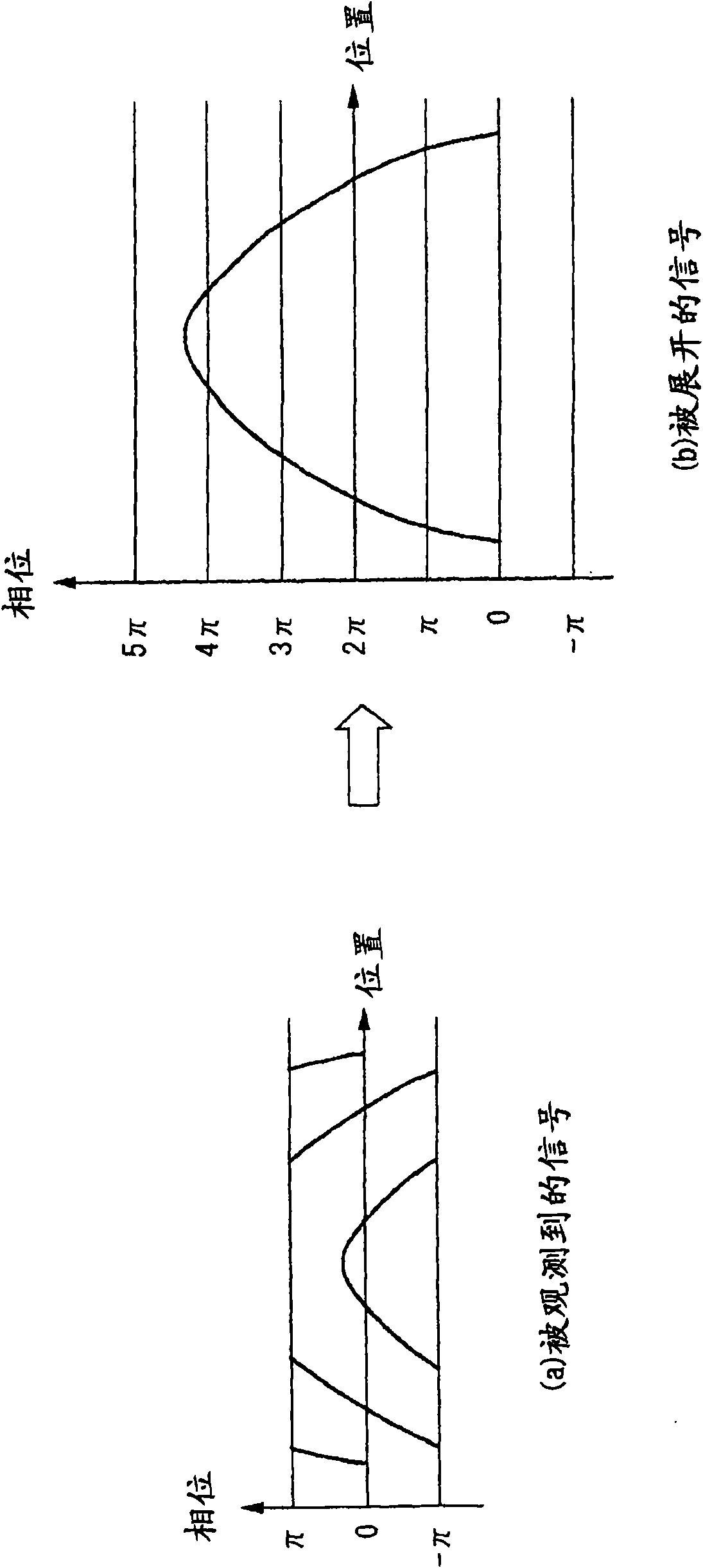
[0034] Ultrasonic diagnostic apparatus 1 transmits and receives ultrasonic waves to a subject such as a living body to measure the moving speed of moving objects such as red blood cells and tissues moving in the subject using phase changes, and displays the measured two-dimensional velocity of the object. Distributed color Doppler ultrasound diagnostic device.

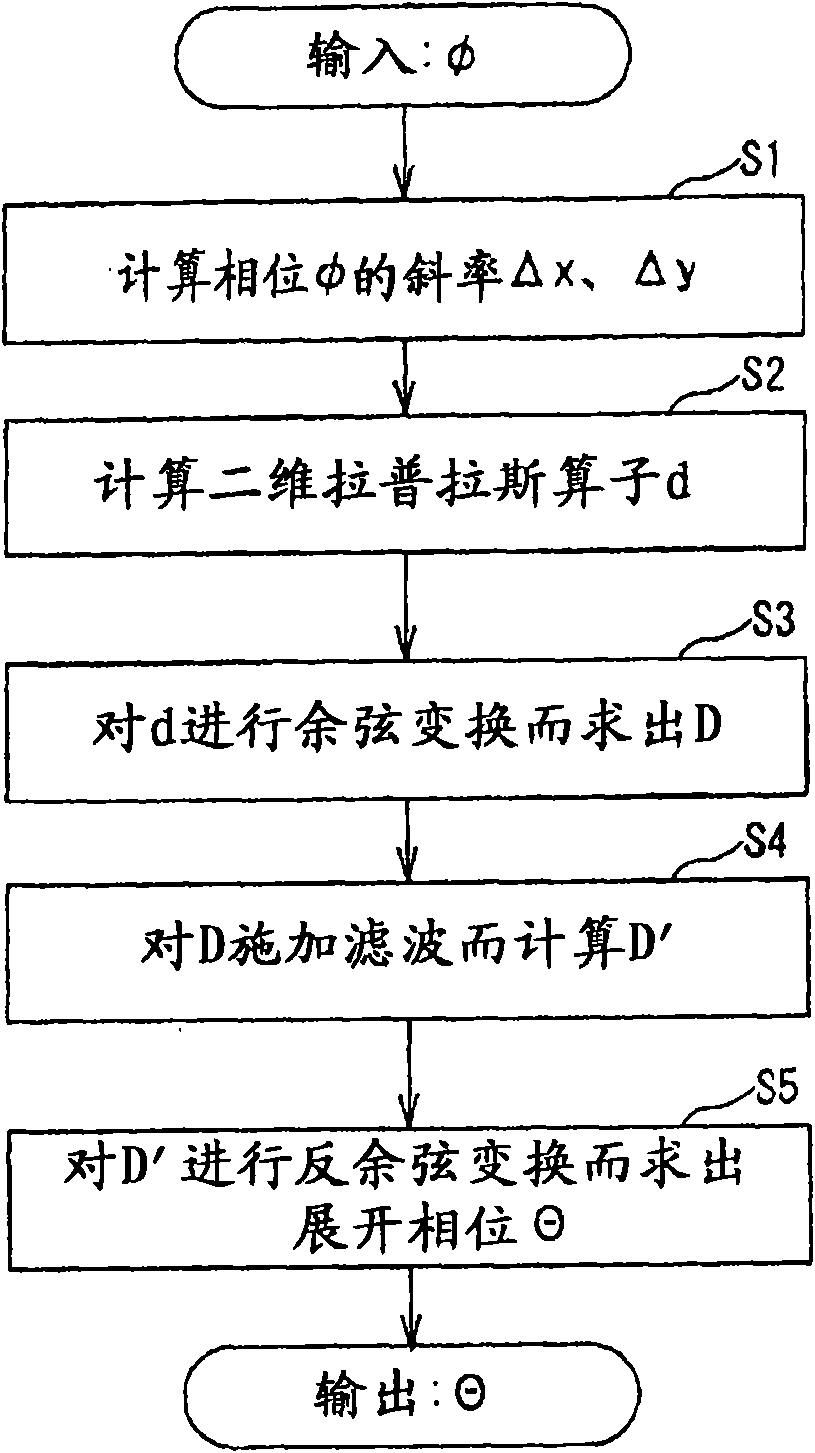
[0035] To this end, the ultrasonic diagnostic apparatus 1 includes a transmitting circuit 2, an ultrasonic probe 3, a receiving circuit 4, a B-mode processing system 5, a color Doppler processing system 6, an aliasi...


PUM


Abstract

An ultrasonographic device includes: a data collection unit which transmits and receives an ultrasonic wave to / from an examinee so as to collect, as Doppler data, a phase signal from a moving body inthe examinee; a correction unit which performs a large-area return correction process on the phase signal in accordance with continuity of a 2-dimensional phase change; and a display unit which displays the phase signal after subjected to the return correction process.

Description

technical field [0001] The present invention relates to an ultrasonic diagnostic apparatus, an ultrasonic diagnostic method, and an ultrasonic diagnostic apparatus for generating and displaying a B-mode image as tissue information of a subject and a color Doppler image as blood flow or tissue motion information by transmitting and receiving ultrasonic waves The data processing program particularly relates to an ultrasonic diagnostic apparatus, an ultrasonic diagnostic method, and a data processing program for an ultrasonic diagnostic apparatus that correct aliasing of velocities in a color Doppler image. Background technique [0002] The color Doppler method in an ultrasonic diagnostic apparatus is a method of irradiating a living body with ultrasonic waves multiple times in the same direction, and extracting blood flow information such as blood flow velocity, energy, and dispersion through the Doppler effect. [0003] In the color Doppler method, a high-pass filter (HPF: hi...

Claims


Application Information

Patent Timeline
Patent Type & Authority Applications(China)
IPC IPC(8): A61B8/06
CPCA61B8/06
Inventor 佐藤武史
Owner TOSHIBA MEDICAL SYST CORP
Features
  • Generate Ideas
  • Intellectual Property
  • Life Sciences
  • Materials
  • Tech Scout
  • Unparalleled Data Quality
  • Higher Quality Content
  • 60% Fewer Hallucinations
Learn More