

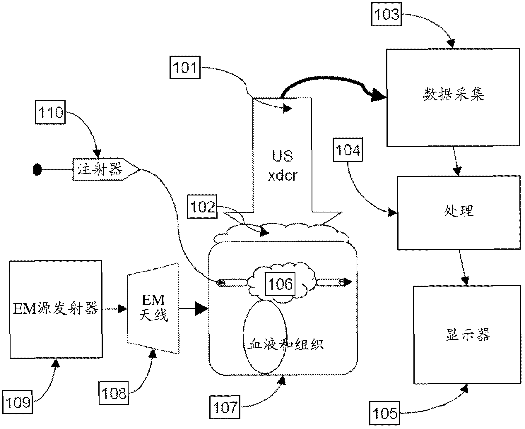
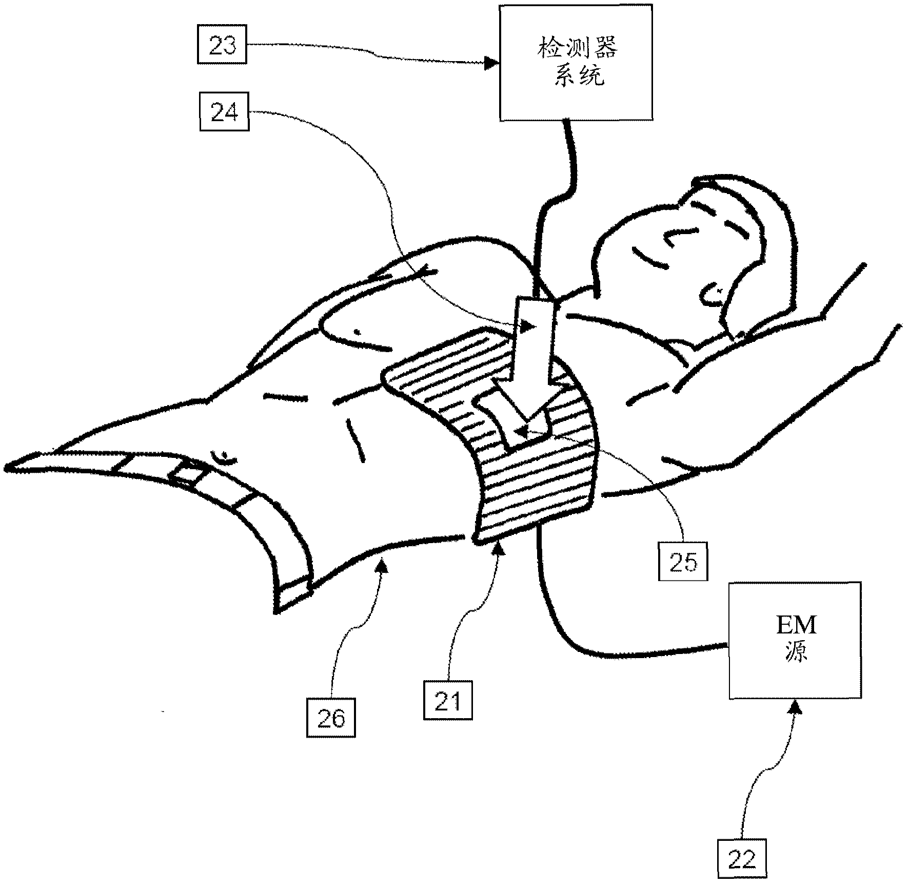
Thermoacoustic system for analyzing tissue
A soft tissue, vasculature technology, used in applications, sonic diagnosis, infrasound diagnosis, etc.
- Summary
- Abstract
- Description
- Claims
- Application Information
AI Technical Summary
Problems solved by technology
Method used
Image

Examples
Embodiment 1
[0084] Example 1. In vitro experiments demonstrating the thermoacoustic approach
[0085] Experiments were performed in vitro to demonstrate the provided thermoacoustic method using a variety of contrast agents. In each experiment, an appropriate contrast agent was placed in a 2-mm tube surrounded by a second aqueous solution and irradiated with pulsed radiofrequency energy, and the resulting thermoacoustic data ( Figure 5 ). Data showed positive (ie increased thermoacoustic signal) and negative (ie decreased thermoacoustic signal) depending on the contrast agent and surrounding medium used in each experiment. Figure 5 The upper left panel of , shows increased signal due to increased ion concentration and thus conductivity and energy absorption in a 2-mm tube of 0.9% saline relative to surrounding deionized water. Figure 5 The upper right panel of , shows the increased signal resulting from irradiation of a 2-mm tube containing 2% saline in an environment of 0.9% saline. ...
Embodiment 2
[0087] Example 2. Spatial resolution of data provided by thermoacoustic methods
[0088] In vitro experiments were performed to determine the spatial resolution of the data provided by the thermoacoustic method. In this experiment, four 0.3-mm tubes containing 5X normal saline (5% NaCl) were placed in a saline environment, the tubes were irradiated with pulsed radiofrequency energy, and the resulting thermoacoustic data ( Figure 6 ) show that thermoacoustic methods can detect deep submillimeter structures with very high contrast.
PUM
 Abstract
Description
Claims
Application Information