Portable radiation imaging system, portable radiation source holder used therein, and set of instruments for radiation imaging

A radiation and portable technology, which is applied in the fields of radiodiagnostic instruments, medical science, tomosynthesis, etc., can solve the problems of handling and setting, the undisclosed X-ray source and system weight, and the reduction of portability, so as to ensure The effect of portability

Inactive Publication Date: 2012-07-11
FUJIFILM CORP
View PDF1 Cites 14 Cited by
  • Summary
  • Abstract
  • Description
  • Claims
  • Application Information

AI Technical Summary

Problems solved by technology

Therefore, a heavy X-ray source will increase the weight of the entire system, causing problems for handling and setup, resulting in reduced portability.
[0006] Even so, US Patent No. 7,664,222 does not disclose the type of X-ray source, nor does it disclose the weight of the X-ray source and system

Method used


Image

  • Portable radiation imaging system, portable radiation source holder used therein, and set of instruments for radiation imaging
  • Portable radiation imaging system, portable radiation source holder used therein, and set of instruments for radiation imaging
  • Portable radiation imaging system, portable radiation source holder used therein, and set of instruments for radiation imaging

Examples


Embodiment Construction

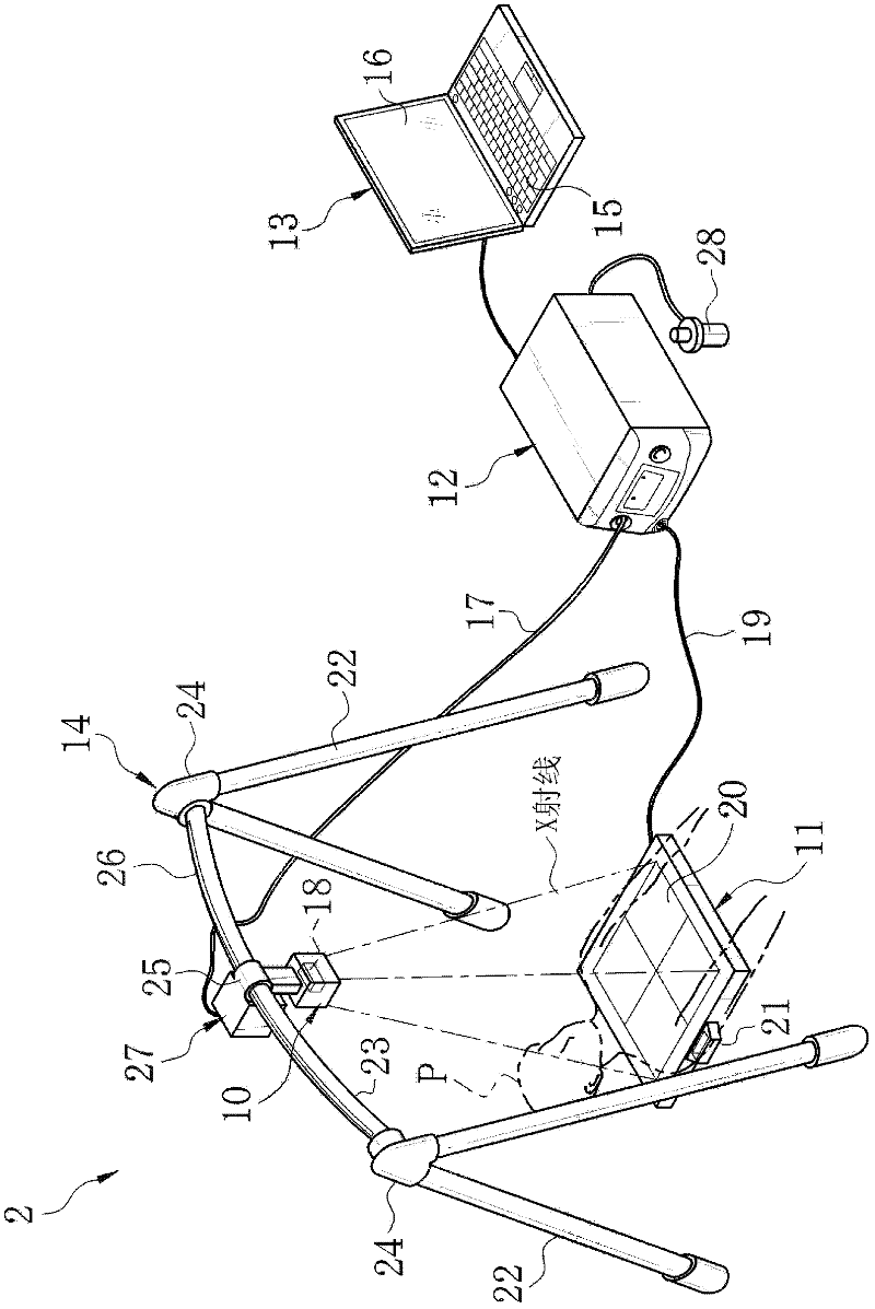
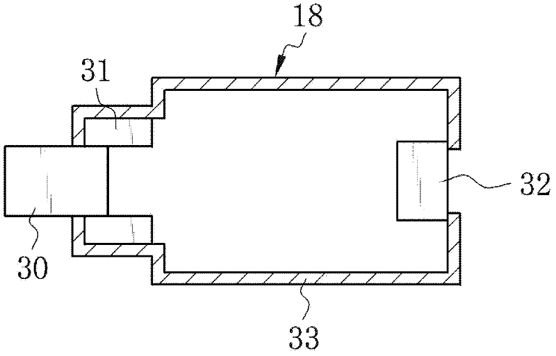
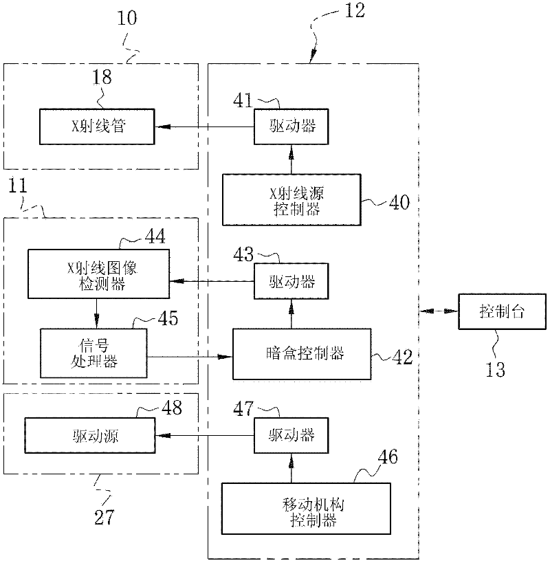
[0025] refer to figure 1 , The X-ray imaging system 2 is composed of an X-ray source 10 , a cassette 11 , an image acquisition control device 12 , a console 13 , and an X-ray source bracket (hereinafter simply referred to as bracket) 14 . The X-ray source 10 emits X-rays to the patient P. As shown in FIG. Cassette 11 detects X-rays emitted from X-ray source 10 and transmitted through patient P, and outputs image data. The image acquisition control device 12 controls the imaging operation of the X-ray source 10 and the cassette 11 . The console 13 sets imaging conditions (tube voltage, tube current, irradiation time, etc. of the X-ray tube 18 of the X-ray source 10 ) for the image acquisition control device 12 . The bracket 14 supports the X-ray source 10 .

[0026] The X-ray source 10, cassette 11, image acquisition control device 12, console 13, and stand 14 are all portable. For X-ray photography, all of these components are brought to a location requiring urgent medical...


PUM


Abstract

In an X-ray imaging system, an X-ray source has a lightweight and small-sized X-ray tube. The X-ray tube is a fixed anode X-ray tube without having a target rotating mechanism. The X-ray tube uses a cold cathode electron source, which does not need a filament and a heater. An image acquisition control device moves the X-ray source to each of plural positions predetermined on a cross bar of a holder by control of a drive source of a shift mechanism. Whenever the X-ray source reaches each position, the X-ray source emits X-rays to an object, and a cassette detects the X-rays transmitted through the object to take an image. Based on data of taken plural images, tomographic image, which puts emphasis on a region of interest inside the object, is produced.

Description

technical field [0001] The present invention relates to a portable radiography system, a radiation source holder and a radiography kit. Background technique [0002] Tomography is well known as a technique for obtaining a tomographic image to precisely examine a region of interest (ROI) inside a human body. In this technique, while moving a radiation source such as an X-ray source (X-ray tube), X-rays are irradiated from the X-ray source to a human body at different angles to capture a plurality of images. The obtained image is processed to generate a tomographic image emphasizing a desired tomographic plane (see US Pat. No. 7,664,222). [0003] US Patent No. 7,664,222 discloses a portable tomography system suitable for use in emergency locations such as the scene of an accident or the scene of a natural or other disaster. The system includes an X-ray source, an X-ray source bracket, an X-ray image detector, a power supply, a control device and a handling device. During t...

Claims


Application Information

Patent Timeline
Patent Type & Authority Applications(China)
IPC IPC(8): A61B6/00
CPCA61B6/107A61B6/025A61B6/4405A61B6/022A61B6/4452A61B6/4283
Inventor 北川祐介西纳直行
Owner FUJIFILM CORP
Why Eureka
Try Eureka