Frequency domain imaging method of ultrasonic scanning microscope

A technology of ultrasonic scanning and imaging methods, which is applied in the analysis of solids by using sonic/ultrasonic/infrasonic waves, and processing the response signal of detection. Effect

Inactive Publication Date: 2013-04-03
BEIJING INSTITUTE OF TECHNOLOGYGY
View PDF8 Cites 10 Cited by
  • Summary
  • Abstract
  • Description
  • Claims
  • Application Information

AI Technical Summary

Problems solved by technology

However, when the frequency of the transducer increases, the attenuation of the sound intensity will increase rapidly, and its penetrating ability will decrease sharply, and generally the higher the frequency, the worse the signal-to-noise ratio, and the echo signal is often buried in the noise signal , the scanned image will instead become blurred due to increased noise

Method used


Image

  • Frequency domain imaging method of ultrasonic scanning microscope

Examples


Embodiment Construction

[0008] The specific embodiment of the present invention is described in detail below:

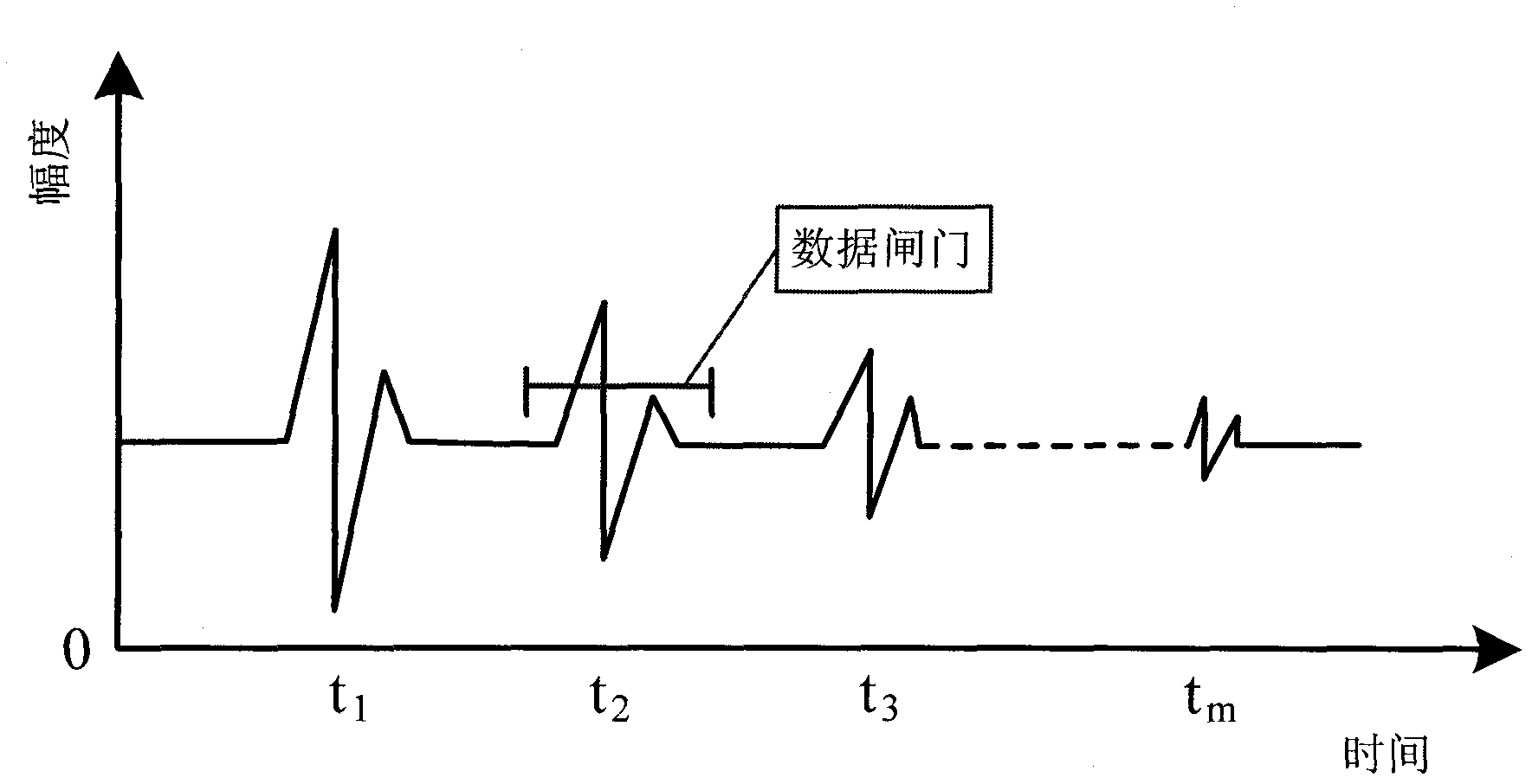
[0009] 1. If figure 1 As shown, adjust the position of the high-frequency focusing transducer in the vertical direction, and observe the echo of the interested part in the A-scan waveform to maximize its amplitude. At this time, the focus of the transducer is placed near this plane position. Move the data gate to jam the echo region that needs to be imaged.

[0010] 2. Set the scanning area, turn on the full-wave acquisition option, perform a C-scan, and save the A-scan full-wave data obtained during the scanning process.

[0011] 3. Open the A-scan full-wave data, perform FFT transformation on the signal in the data gate area of ​​each A-scan waveform in turn, and then find the value with the largest amplitude in the obtained spectrum amplitude, and record it in a two-dimensional array.

[0012] 4. After the FFT transformation of A-scan waveforms in all incident directions is completed, ...


PUM


Abstract

The invention relates to a frequency domain imaging method of an ultrasonic scanning microscope, being applicable to the field of ultrasonic scanning microscope detection and ultrasonic C-scanning detection and capable of improving the C-scanning imaging precision in ultrasonic micro-scanning and revealing details which are difficultly discovered in time-domain imaging. The frequency domain imaging method comprises the following steps of: firstly, carrying out full-wave acquisition on an A-scanning signal generated in the C-scanning process; secondly, carrying out fast Fourier transform on signals in data gates of each A-scanning waveform, finding out the maximum amplitudes in the signals, normalizing the maximum amplitudes and expressing the intensities by using corresponding color gray levels; and finally, drawing pixel points on a screen so as to accomplish the frequency domain imaging in ultrasonic micro-scanning.

Description

1. Technical field [0001] The invention relates to a frequency-domain imaging method of an ultrasonic scanning microscope, which is suitable for the fields of ultrasonic scanning microscope detection and ultrasonic C-scan detection. 2. Background technology [0002] Ultrasonic scanning microscope is a very effective means to detect internal defects in electronic packaging structures. It is a method that uses focused high-frequency ultrasound, usually 10MHz to 2GHz, to visualize the surface, subsurface, and fine structures within a certain depth of the object. Micro-imaging is a technology for visual observation. It is mainly aimed at the failure analysis of semiconductor devices, chips, and materials. It can check the lattice structure inside the material, impurity particles, internal cracks, layered defects, voids, bubbles, etc. [0003] The high-frequency focused ultrasonic transducer used in the ultrasonic scanning microscope is the most critical component, and its select...

Claims


Application Information

Patent Timeline
Patent Type & Authority Applications(China)
IPC IPC(8): G01N29/06G01N29/46
Inventor 徐春广刘中柱门伯龙赵新玉肖定国郭祥辉杨柳李喜朋阎红娟
Owner BEIJING INSTITUTE OF TECHNOLOGYGY
Why Eureka
Try Eureka