Reagent device and method for detecting anti-mitochondrial antibodies type M2 antibody

An anti-mitochondrial antibody and detection reagent technology, applied in the field of clinical immunology detection, can solve the problems such as the need to improve the sensitivity, poor accuracy and precision of the detection result, complicated operation, etc. Apply a wide range of effects

Active Publication Date: 2013-07-03
SHENZHEN YHLO BIOTECH
View PDF6 Cites 3 Cited by
  • Summary
  • Abstract
  • Description
  • Claims
  • Application Information

AI Technical Summary

Problems solved by technology

[0007] (1) Non-specific recognition cannot be distinguished according to the size of the molecular weight when analyzing the results;
[0008] (2) The operation is relatively complicated and requires expensive fluorescence microscopes, which are difficult to popularize in many primary hospitals and are not suitable for laboratories with a large number of specimens;
[0009] (3) The background in the fluorescence measurement is relatively high, and it is difficult to use the fluorescence immunoassay technique for quantitative determination;
[0010] (4) Experienced professionals are required to determine the results, and the objectivity of the analysis results is insufficient;
[0011] (5) Only qualitative detection can be carried out, and quantitative measurement cannot be carried out
[0014] (1) Only qualitative and semi-quantitative analysis can be carried out, and the specific amount of the tested substance cannot be obtained
[0015] (2) The operation steps are cumbersome and the test takes a long time
[0016] (3) The detection sensitivity needs to be improved
[0019] (1) Use 12×8 type, 6×8 type, 8×12 type or whole plate type 96-well special microwell plate as the antigen coating equipment and reaction container, which can only be divided into 12 batches and 6 batches when used , 8 batches or the whole board can be used at one time, and independent and single-person testing cannot be carried out;
[0020] (2) There are many kinds of reagents used in quantitative determination, and each detection reagent must be contained in a reagent b

Method used


Image

  • Reagent device and method for detecting anti-mitochondrial antibodies type M2 antibody
  • Reagent device and method for detecting anti-mitochondrial antibodies type M2 antibody
  • Reagent device and method for detecting anti-mitochondrial antibodies type M2 antibody

Examples


Example Embodiment

[0095] Example 1 Reagent device 1 for detecting anti-mitochondrial antibody M2 type

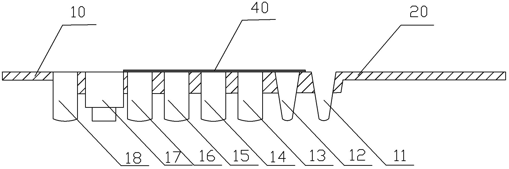
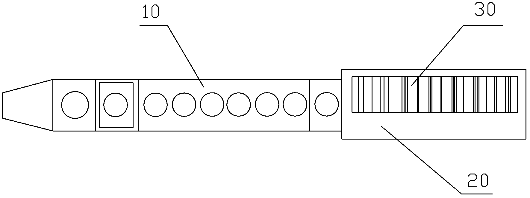
[0096] Such as Figure 1-2 As shown, the reagent device for detecting anti-mitochondrial antibody M2 type described in the application of the present invention is a long strip, including a base 10 with eight holes and a handle 20 at one end of the base 10. The eight holes are arranged in order from the nearest The end of the handle 10 is followed by sample hole 11, adjuvant hole 12, enzyme conjugate hole 13, substrate hole 14, stop solution hole 15, diluent hole 16, reaction hole 17 and dilution hole 18. The sample hole 11 The sample to be tested is contained in the auxiliary agent hole 12 for adding auxiliary reagents when needed for detection. In the detection method described in the present application, no reagent is added to the auxiliary agent hole 12, and the enzyme conjugate is added to the enzyme conjugate hole 13 Solution, the substrate solution is added to the substrate hole 14, the st...

Example Embodiment

[0099] Example 2 Reagent device 2 for detecting anti-mitochondrial antibody M2 type

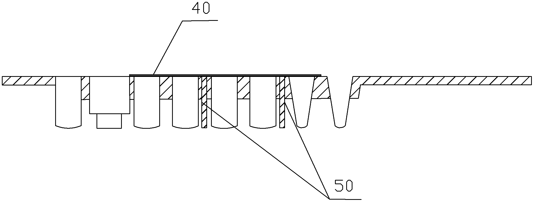
[0100] Such as image 3 As shown, the basic structure of the reagent device for detecting anti-mitochondrial antibody M2 type in this embodiment is the same as that of the reagent device in the first embodiment. The reagent device also includes several support columns 50. Located below the substrate 10 in the reagent device, there are more than one hole positions between adjacent support columns 50. The function of the support columns 50 is to enhance the mechanical strength and balance of the substrate.

Example Embodiment

[0101] Example 3 Reagent device 3 for detecting anti-mitochondrial antibody M2 type

[0102] Such as Figure 4-6 Shown is a preferred embodiment of the reagent device for detecting anti-mitochondrial antibody M2 type according to the application of the present invention. Its basic structure is the same as that of Embodiment 1 or Embodiment 2, except that the reaction hole 17 is a detachable structure. The hole 17 is composed of an outer hole 171 and an inner hole 172. A bottom hole 174 is opened at the bottom of the outer hole 171. The inner hole 172 passes through the bottom hole 174 and is tightly fitted with the bottom hole 174. The anti-overflow cavity 173, the function of the anti-overflow cavity 173 is to stay in the cavity if the liquid overflows during the reaction process to prevent contamination of the instrument and other reagent devices.

[0103] Further, the method of cooperating and fixing the inner hole and the outer hole can also be to design the outer wall of the ...


PUM


Abstract

The invention provides a reagent device and a method used for detecting anti-mitochondrial antibodies type M2 antibodies. The reagent device has a long strip shape, and has a base body comprising 8 hole positions and a handle positioned on one end of the base body. From the end proximal to the handle, the 8 hole positions are sequentially a sample hole, an auxiliary agent hole, an enzyme conjugate hole, a substrate hole, a termination liquid hole, a dilution liquid hole, a reaction hole, and a dilution hole. According to the invention, based on a principle of enzyme-linked immunoassay, anti-mitochondrial antibodies type M2 antibody is detected by using the reagent device. The method is an independent single-person analysis and detection method. The device can be used in cooperation with a corresponding specific analysis instrument. During a detection process, detection reagents or samples are injected by using a full-automatic precise dosing device. The device and the method have the advantages of automatic operation, precise dosing, high detection result accuracy, high detection result precision, and wide application prospect.

Description

technical field [0001] The application of the present invention relates to a reagent device and a method for detecting the M2 type of anti-mitochondrial antibody, belonging to the technical field of clinical immunology detection. Background technique [0002] Primary biliary cirrhosis (PBC) is a chronic progressive disease, which is more common in women. It is due to the progressive destruction of small and medium bile ducts in the liver accompanied by inflammatory changes in the portal vein, and further develops into fibrosis and cirrhosis. , clinically manifested as progressive liver dysfunction. In the early stage, it manifests as skin itching, lethargy, and dull pain in the liver area, and in the late stage, jaundice, hepatic ascites, and liver cirrhosis appear. PBC is an autoimmune disease, and 80% of patients are accompanied by other immune diseases. Anti-mitochondrial (AMA) antibody M2 type, anti-nuclear antibody, anti-nuclear pore complex antibody and other autoant...

Claims


Application Information

Patent Timeline
IPC IPC(8): G01N33/543G01N21/31
Inventor 胡德明刘清波何林阳辉
Owner SHENZHEN YHLO BIOTECH
Why Eureka
Try Eureka