Three-dimensional color coding display method for myocardial blood volume distribution

A technology for color-coding and displaying methods for applications in diagnostic recording/measurement, medical science, sensors, etc.

Inactive Publication Date: 2014-03-19
UNIV OF SCI & TECH OF CHINA
View PDF6 Cites 4 Cited by
  • Summary
  • Abstract
  • Description
  • Claims
  • Application Information

AI Technical Summary

Problems solved by technology

The electrocardiogram contains information on the distribution of myocardial blood volume in the heart, but it is not a two-dimensional or

Method used


Image

  • Three-dimensional color coding display method for myocardial blood volume distribution
  • Three-dimensional color coding display method for myocardial blood volume distribution
  • Three-dimensional color coding display method for myocardial blood volume distribution

Examples


Embodiment Construction

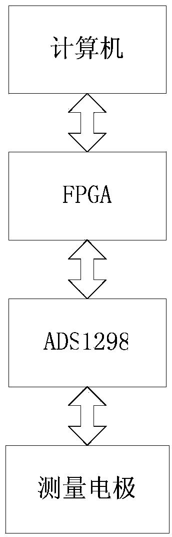
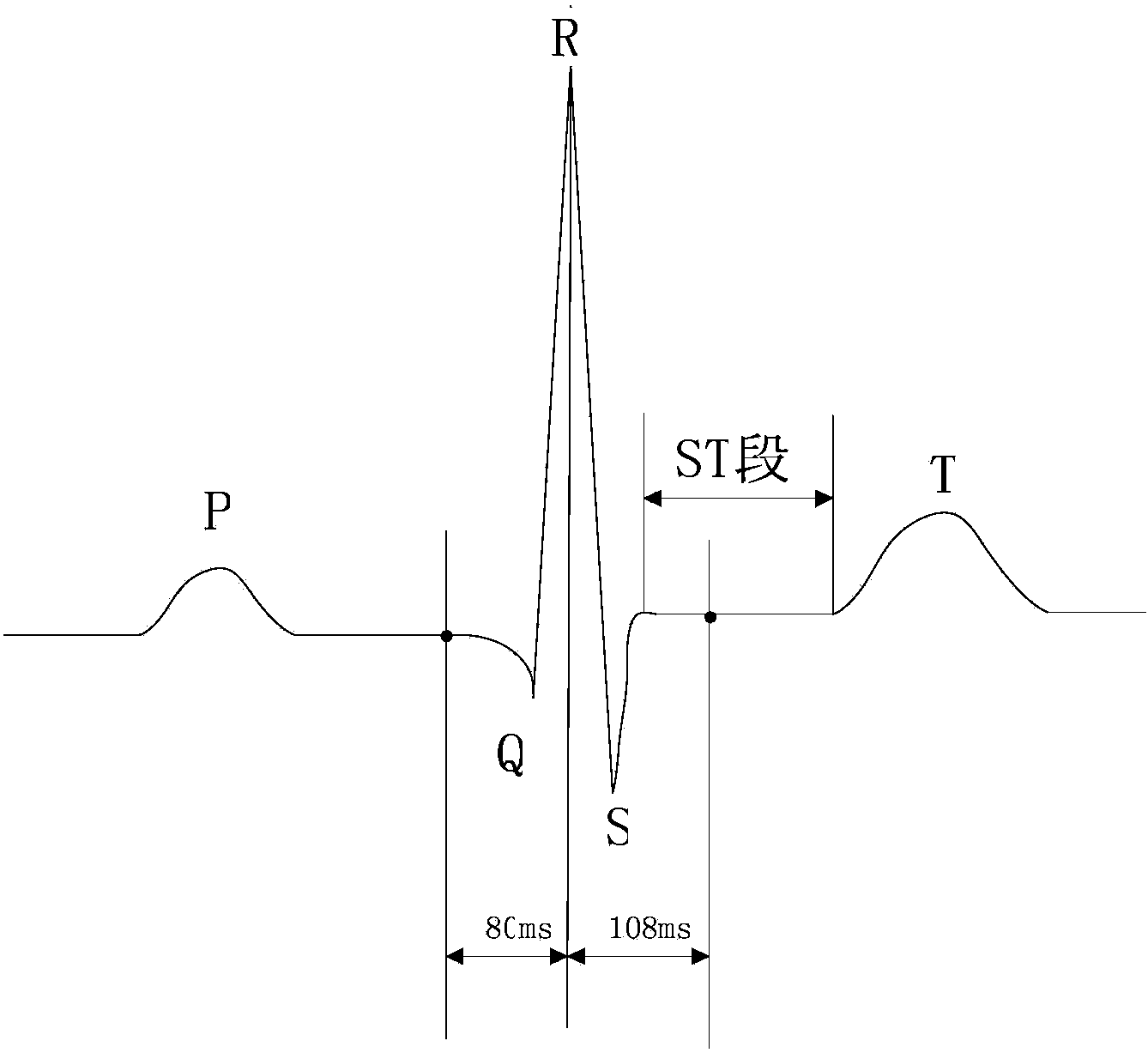
[0018] figure 1 The hardware block diagram of ECG signal collection is described. The ECG signal collected by electrodes is converted into digital signal by using ADS1298. The digital signal is processed by FPGA and transmitted back to the computer. The sampling rate is 500SPS. The computer controls the working mode of ADS1298 through the host computer program.

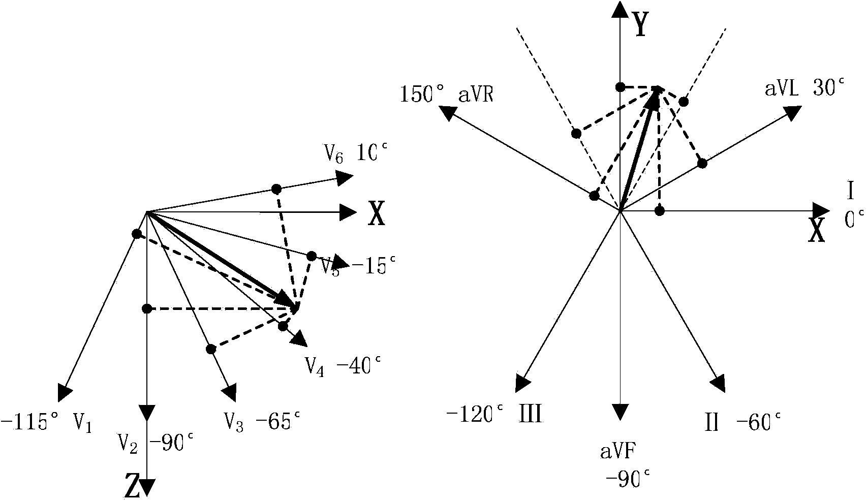
[0019] The electrode connection method is as follows: lead V1 is at the 4th intercostal space on the right border of the sternum, lead V2 is at the 4th intercostal space at the left border of the sternum, lead V3 is at the midpoint of the line connecting V2 and V4, and lead V4 is at the left midclavicular line and the 5th intercostal space, V 5 lead and V 6 lead with V 4 The leads are at the same level, located at the left anterior axillary line and the left midaxillary line respectively. The lead wires are red for the right upper extremity, yellow for the left upper extremity, green for the left lower extremity, ...


PUM


Abstract

The invention relates to a three-dimensional color coding display method for myocardial blood volume distribution, through which the blood volume distribution information of electrocardiosignals is converted and displayed on a visual three-dimensional color model. The method is characterized in that a 12-lead electrocardiogram (ECG) of a human body is collected, and the myocardial blood volume distribution information of the ECG, i.e. ST-segment deviation, is extracted; in a cardiac electric axis system, the total electrocardial damage vector is obtained through the adverse projection of the ST-segment damage vector of each lead; a standard three-dimensional heart model is established, the central position of an area where the blood volume is scarcely distributed is the joint of the total damage vector and the surface of the heart model, the distribution range is proportional to the total damage vector, and RGB (red, green and blue) coding is carried out according to the different distances between all the positions and the center of the area where the blood volume is scarcely distributed, so as to display the myocardial blood volume distribution on the surface of the heart model.

Description

technical field [0001] The invention relates to a method for displaying myocardial blood volume distribution through an intuitive three-dimensional color heart model, in particular to a display method for converting one-dimensional electrocardiographic signals into three-dimensional color codes. Background technique [0002] Electrocardiogram: In each cardiac cycle, the heart is successively excited by the pacemaker, atrium, and ventricle, accompanied by changes in bioelectricity, and various forms of potential changes are drawn from the body surface through the electrocardiograph (ECG for short). The electrocardiogram is an objective indicator of the occurrence, propagation and recovery process of cardiac excitement. The electrocardiogram contains information on the distribution of myocardial blood volume in the heart, but it is not a two-dimensional or three-dimensional blood volume distribution. The pure one-dimensional electrocardiogram signal can only analyze the distri...

Claims


Application Information

Patent Timeline
IPC IPC(8): A61B5/0402
Inventor 蒋依秦杨圣蔡莎莎张韶宇
Owner UNIV OF SCI & TECH OF CHINA
Why Eureka
Try Eureka