
A medical image scanning method and device

A technology of medical images and scanning methods, which is applied in computer tomography scanners, echo tomography, etc., can solve the problems of inability to judge the scanning position, unintuitive modification, limited follow-up 3D and advanced application functions, etc., to achieve completeness Effect

Active Publication Date: 2018-05-04
SHANGHAI UNITED IMAGING HEALTHCARE
View PDF10 Cites 0 Cited by
  • Summary
  • Abstract
  • Description
  • Claims
  • Application Information

AI Technical Summary

Problems solved by technology

Using the parameter box to directly edit the settings can strictly ensure that the start position of the supplementary scan is the end position of the previous scan, but the modification is not intuitive enough, and the user cannot accurately judge the scan position
Dragging and dropping settings through the positioning box generally cannot guarantee that the start position of the supplementary scan is the end position of the previous scan, resulting in that the original data and images obtained by the supplementary scan cannot be directly merged with the original sequence, and the functions of subsequent 3D and advanced applications are affected. limit

Method used


Image

  • A medical image scanning method and device
  • A medical image scanning method and device

Examples


Embodiment Construction

[0029] The present invention will be further described below in conjunction with the accompanying drawings and embodiments.

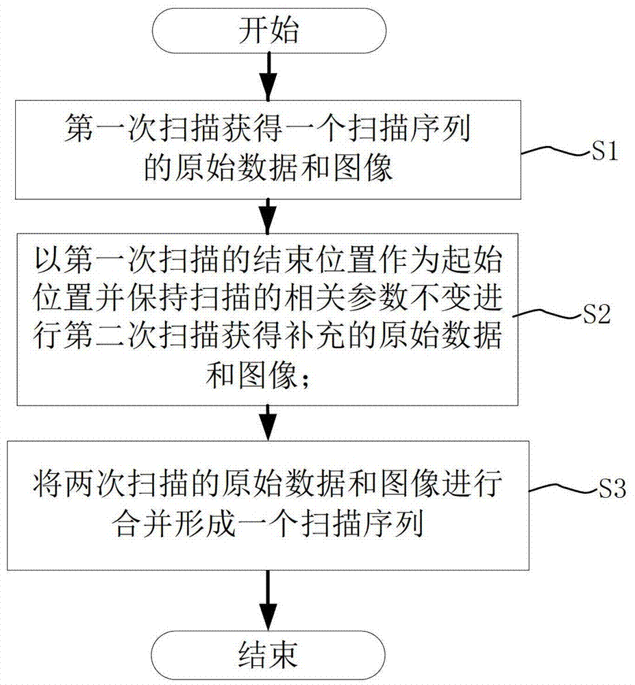
[0030] figure 1 It is a flowchart of medical image scanning in the present invention.

[0031] See figure 1 , the medical image scanning method provided by the present invention comprises the following steps:

[0032] Step S1: Edit directly through the parameter box or set the start position, scan length and end position through the positioning box, and obtain the original data and image of a scan sequence in the first scan; the image is the image obtained after reconstruction based on the original data .

[0033] Step S2: Take the end position of the first scan as the starting position and keep the scanning and reconstruction parameters unchanged for the second scan to obtain supplementary raw data and images; that is, set the end position of the first scan as the starting point of the supplementary scan The initial position, and keep the scanning ...


PUM


Abstract

The invention discloses a medical image scanning method and device. The medical image scanning method includes the following steps: a) obtaining the original data and images of a scanning sequence by the first scan; b) taking the end position of the first scan as the Starting position and keeping the relevant parameters of scanning and reconstruction unchanged, perform a second scan to obtain supplementary raw data and images; c) combine the raw data and images obtained from the first scan and the second scan to form a scan sequence. The medical image scanning method and device provided by the present invention take the end position of the first scan as the start position of the second scan and keep the image related parameters unchanged to perform fast and accurate supplementary scans, and synthesize a sequence with the original sequence , so as to ensure the integrity of the scanning sequence and facilitate subsequent processing.

Description

technical field [0001] The invention relates to an image scanning method and device, in particular to a medical image scanning method and device. Background technique [0002] When scanning medical images, if there is a deviation during the scanning process, it is often necessary to perform a supplementary scan. At present, the method of supplementary scanning of medical images is relatively simple, mainly through the direct editing and setting method of the parameter box or dragging and dropping setting through the positioning box. Since there can only be one fixed scanning direction during scanning, the starting position of the supplementary scan is actually the end position of the previous scan, so that the original data and images of the supplementary scan can be merged with the original data and images of the previous scan. Although using the parameter box to directly edit the settings can strictly ensure that the start position of the supplementary scan is the end pos...

Claims


Application Information

Patent Timeline
Patent Type & Authority Patents(China)
IPC IPC(8): A61B6/03
Inventor 吴晓明
Owner SHANGHAI UNITED IMAGING HEALTHCARE
Learn More