Positioning method of multiple organs in three-dimensional image

A technology of three-dimensional image and positioning method, which is applied in the field of image processing, and can solve the problems of generalized Hough transform prone to positioning errors, etc.

Active Publication Date: 2014-10-29
SHANGHAI UNITED IMAGING HEALTHCARE
View PDF3 Cites 6 Cited by
  • Summary
  • Abstract
  • Description
  • Claims
  • Application Information

AI Technical Summary

Problems solved by technology

However, it is worth noting that the generalized Hough transform currently used in organ localization only uses the morphological model of the organ to be localized, and discards the important cont

Method used


Image

  • Positioning method of multiple organs in three-dimensional image
  • Positioning method of multiple organs in three-dimensional image
  • Positioning method of multiple organs in three-dimensional image

Examples


Embodiment Construction

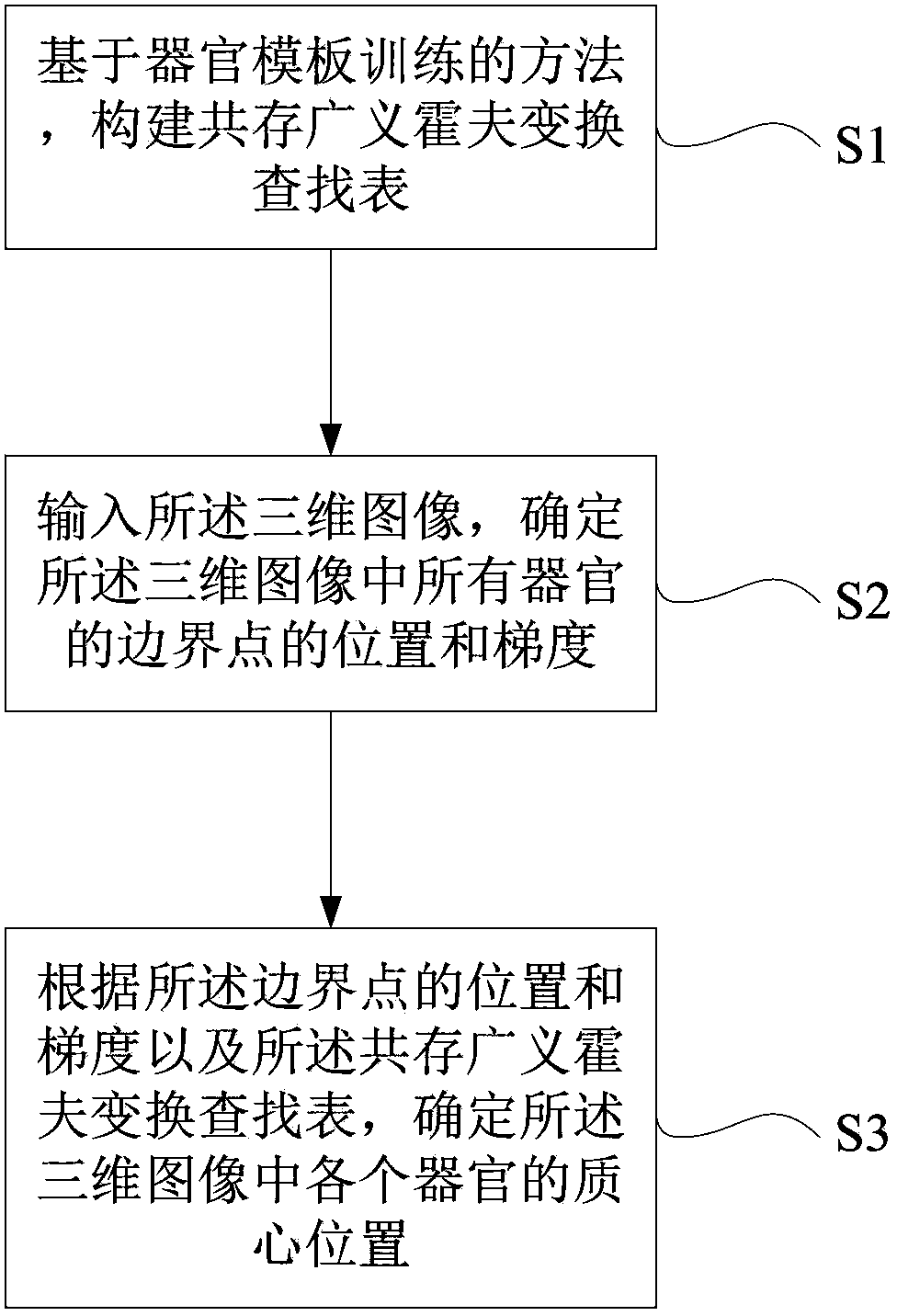
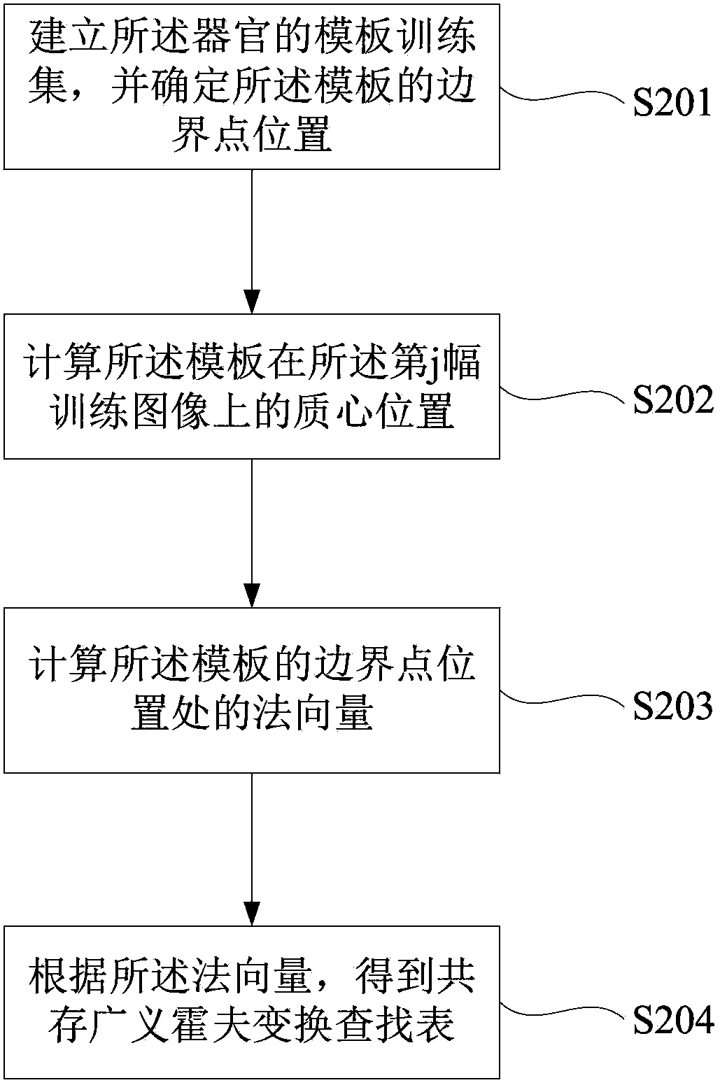
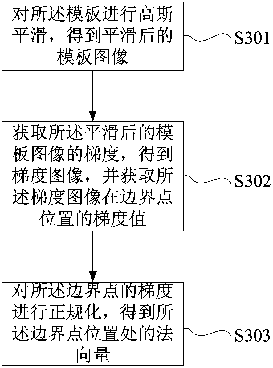
[0063] In the following description, numerous specific details are set forth in order to provide a thorough understanding of the present invention. However, the present invention can be implemented in many other ways different from those described here, and those skilled in the art can make similar extensions without violating the connotation of the present invention, so the present invention is not limited by the specific implementations disclosed below.

[0064] Secondly, the present invention is described in detail by means of schematic diagrams. When describing the embodiments of the present invention in detail, for convenience of explanation, the schematic diagrams are only examples, which should not limit the protection scope of the present invention.

[0065] The present invention will be described in detail below in conjunction with the accompanying drawings and embodiments. The positioning method of multiple organs in the three-dimensional image of the present inventi...


PUM


Abstract

The invention provides a positioning method of multiple organs in a three-dimensional image. The positioning method comprises constructing a coexistence generalized Hough transform lookup table of the offset of a boundary point of any one organ relative to the positions of the center of mass of all the organs based on an organ template training method; inputting a three-dimensional image, and determining positions and gradients of the boundary points of all the organs in the three-dimensional image; and based on the positions and gradients of the boundary points and the lookup table, determining the position of the center of mass of each organ in the three-dimensional image, that is determining the position of each organ in the three-dimensional image. According to the invention, by use of the form information on an organ in need of position and relative position relation information with other organs, the position of the organ in the image can be quickly, accurately and stably positioned.

Description

technical field [0001] The invention relates to the field of image processing, in particular to a multi-organ positioning method in a three-dimensional image. Background technique [0002] Fast, accurate and stable localization of human organs in medical images is usually an important step in computer-aided diagnosis. The automatic segmentation of organs in medical images, the extraction and quantitative analysis of organ structures (including lesions) all depend on accurately locating the location of each organ in complex medical images. [0003] Organ localization methods in medical images can be roughly classified into two categories. The first type of method usually does not require any prior organ model, but searches a large number of medical images as training samples, extracts the features of each organ in the image through machine learning, and determines the organ of interest based on these features Position in an unknown image [S.Seifert, A.Barbu, S.K.Zhou, D.Liu...

Claims


Application Information

Patent Timeline
IPC IPC(8): G06T7/00
Inventor 刘靖孙贤军庄吓海李强
Owner SHANGHAI UNITED IMAGING HEALTHCARE
Why Eureka
Try Eureka