
Mesenchymal stem cells from meniscus tissues as well as preparation method and identification thereof

A mesenchymal stem cell and meniscus technology, applied in the field of mesenchymal stem cells and its preparation, can solve the problems of limited number of meniscus cells, inaccuracy of meniscus cells, poor repair effect, etc., and achieve good clinical application prospects

Inactive Publication Date: 2015-02-18
WEST CHINA HOSPITAL SICHUAN UNIV
View PDF2 Cites 0 Cited by
  • Summary
  • Abstract
  • Description
  • Claims
  • Application Information

AI Technical Summary

Problems solved by technology

[0004] The terminally differentiated mature autologous meniscus cells are derived from the injured meniscus tissue, which are derived from autologous and exact meniscus cell phenotype characteristics, and have always been considered as the most ideal seed cells for meniscus regeneration; however, they are also used as seed cells. There are some problems: two operations are required, the number of meniscus cells derived from diseased tissue is very limited, and the lack of proliferation ability and biological activity of normal meniscus cells, etc.
Although there are many reports on the use of mesenchymal stem cells from other tissues, such as adipose or synovial tissue, as seed cells for meniscus regeneration, the biggest disadvantage of these tissue-derived mesenchymal stem cells is their inability to differentiate into meniscus cells. exactness, leading to poor repair results
[0006] To sum up, the seed cells currently used for meniscal tissue repair are either insufficient in "stemness" or difficult to effectively differentiate into meniscus cells, resulting in poor repair effect

Method used


Image

  • Mesenchymal stem cells from meniscus tissues as well as preparation method and identification thereof
  • Mesenchymal stem cells from meniscus tissues as well as preparation method and identification thereof
  • Mesenchymal stem cells from meniscus tissues as well as preparation method and identification thereof

Examples


Embodiment 1

[0033] Example 1 Isolation and detection of mesenchymal stem cells derived from meniscus tissue of the present invention

[0034] 1. Separation and detection method

[0035] 1. Collection of meniscus fragments

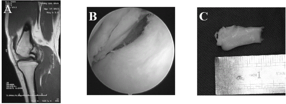
[0036] The diagnosis of meniscus injury was diagnosed comprehensively based on the patient's clinical manifestations, positive findings under MRI and arthroscopy (see figure 1 ). Arthroscopically removed meniscal fragments with nucleus forceps (see figure 1 ). The tissues with blood vessels, the surrounding synovium and ligaments were excised under a microscope, and rinsed 3 times in PBS containing double antibodies.

[0037] 2. Isolation and culture of tissue-specific mesenchymal stem cells derived from meniscus fragments

[0038] Use ophthalmic scissors to cut the collected meniscus fragments into about 1-5mm 3 Tissue fragments of different sizes were then shaken in a 0.2% type I, 0.2% type II mixed collagenase (concentrations of both type I and type II collage...

Embodiment 2

[0039] Example 2 Isolation and detection of mesenchymal stem cells derived from meniscus tissue of the present invention

[0040]1. Separation and detection method

[0041] 1. Collection of meniscus fragments

[0042] The diagnosis of meniscus injury was diagnosed comprehensively based on the patient's clinical manifestations, positive findings under MRI and arthroscopy (see figure 1 ). Arthroscopically removed meniscal fragments with nucleus forceps (see figure 1 ). The tissues with blood vessels, the surrounding synovium and ligaments were excised under a microscope, and rinsed 3 times in PBS containing double antibodies.

[0043] 2. Isolation and culture of tissue-specific mesenchymal stem cells derived from meniscus fragments

[0044] Use ophthalmic scissors to cut the collected meniscus fragments into about 1-5mm 3 Tissue fragments of different sizes were then shaken in a 0.1% type I and 0.1% type II mixed collagenase (concentrations of both type I and type II colla...

Embodiment 3

[0045] Example 3 Isolation and detection of mesenchymal stem cells derived from meniscus tissue of the present invention

[0046] 1. Separation and detection method

[0047] 1. Collection of meniscus fragments

[0048] The diagnosis of meniscus injury was diagnosed comprehensively based on the patient's clinical manifestations, positive findings under MRI and arthroscopy (see figure 1 ). Arthroscopically removed meniscal fragments with nucleus forceps (see figure 1 ). The tissues with blood vessels, the surrounding synovium and ligaments were excised under a microscope, and rinsed 3 times in PBS containing double antibodies.

[0049] 2. Isolation and culture of tissue-specific mesenchymal stem cells derived from meniscus fragments

[0050] Use ophthalmic scissors to cut the collected meniscus fragments into about 1-5mm 3 Tissue fragments of different sizes were then shaken in a 0.5% type I, 0.5% type II mixed collagenase (concentrations of both type I and type II collage...


PUM


Abstract

The invention discloses a method for separating mesenchymal stem cells from meniscus tissues. The method comprises the following steps: (1) shearing a meniscus tissue into tissue fragments, and digesting for 4-6 hours by a mixed collagenase solution containing I-type collagenase and II-type collagenase; (2) terminating digestion, filtering by a 100-400mesh screen, centrifuging for 4-10 minutes at 1000-2000rpm, removing supernate, washing for 3 times by PBS, re-suspending in a mesenchymal stem culture medium, culturing at 37 DEG C and 5% CO2, removing non-adherent cells and fragments after 2-3 days, changing fluid every 3 days, and harvesting cells when the fusion degree is 80-90 percent. According to the method, the mesenchymal stem cells obtained by separation integrate the advantages of stable phenotype of meniscus cells and 'stemness' characteristic of the mesenchymal stem cells, have exact differentiation and excellent bioactivity, are a relatively ideal seed cell source in meniscus regeneration and clinical translational research thereof in the future, and have excellent clinical application prospect.

Description

technical field [0001] The invention relates to a mesenchymal stem cell derived from meniscus tissue, a preparation method and identification thereof. Background technique [0002] The meniscus plays very important biomechanical and biochemical functions in maintaining the homeostasis of the knee joint, including absorbing shock, transmitting load, stabilizing the joint, dispersing stress, and exerting the proprioception of the knee joint. Meniscal injury is a very common sports- and age-related lesion in clinic. Damage to anatomical structures will lead to joint degeneration and osteoarthritis, accompanied by varying degrees of joint dysfunction. The treatment of meniscus injury has always been a difficult problem in the basic research and clinical practice of orthopedics and sports medicine. The current clinical treatment methods for meniscus injury generally include meniscectomy, suture repair, and reconstruction. Removal of part or all of the meniscus will result in i...

Claims


Application Information

Patent Timeline
Patent Type & Authority Applications(China)
IPC IPC(8): C12N5/0775
Inventor 付维力
Owner WEST CHINA HOSPITAL SICHUAN UNIV
Learn More