
Image processing device, method, and program

An image processing device and image technology, which is applied in image data processing, image data processing, image memory management, etc., can solve the problem that the accuracy of region extraction is not so good

Inactive Publication Date: 2015-06-17
FUJIFILM CORP
View PDF5 Cites 0 Cited by
  • Summary
  • Abstract
  • Description
  • Claims
  • Application Information

AI Technical Summary

Problems solved by technology

[0004] However, the method described in Patent Document 1 is to extract a region from a two-dimensional image, so the extraction range may be subtly different in each two-dimensional image, so the extraction accuracy of the region in the case of being regarded as a three-dimensional image is not so it is good

Method used


Image

  • Image processing device, method, and program
  • Image processing device, method, and program
  • Image processing device, method, and program

Examples


Embodiment Construction

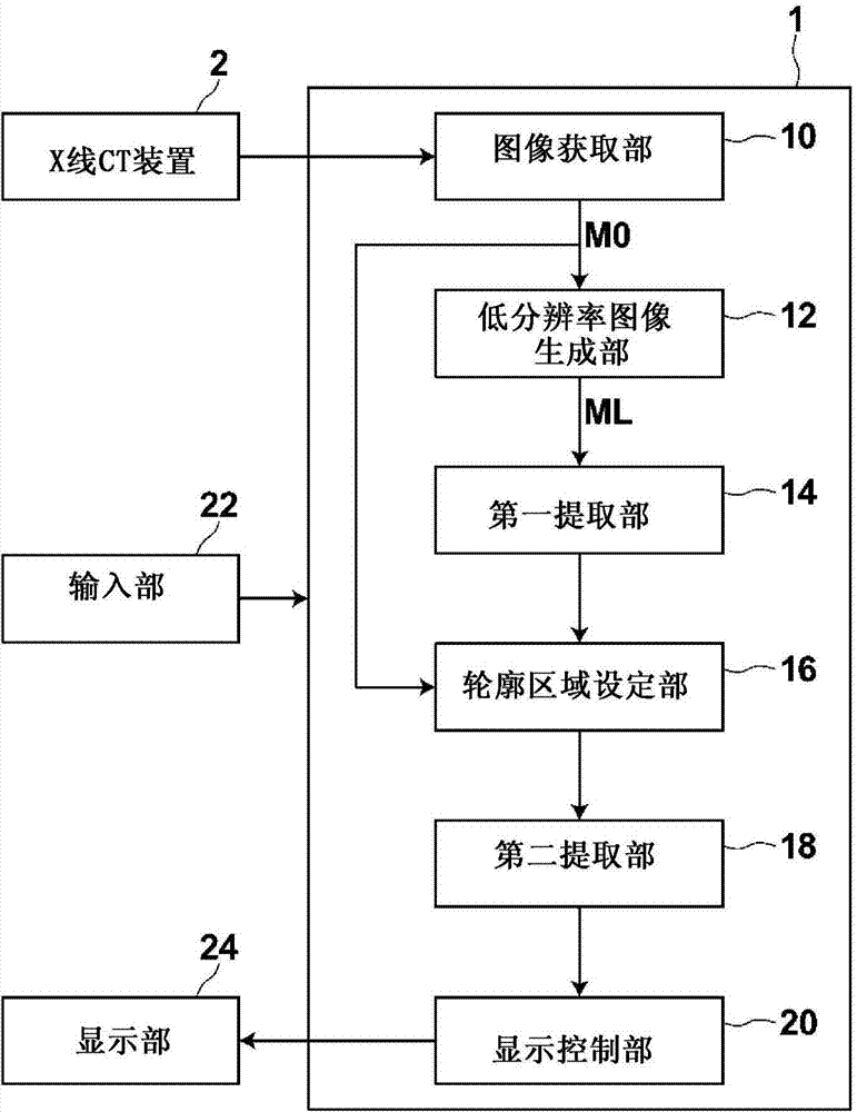
[0039] Hereinafter, embodiments of the present invention will be described with reference to the drawings. figure 1 It is a schematic block diagram showing the configuration of the image processing device according to the embodiment of the present invention. also, figure 1 The configuration of the shown image processing apparatus 1 is realized by executing a program read into an auxiliary storage device (not shown) on a computer (for example, a personal computer, etc.). In addition, this program is stored in an information storage medium such as a CD-ROM, or distributed via a network such as the Internet, and installed in a computer.

[0040] The image processing device 1, for example, generates a three-dimensional image M0 from a plurality of two-dimensional images captured by the X-ray CT apparatus 2, automatically extracts a specific region contained in the three-dimensional image M0 using a graph cut method, and includes: an image acquisition unit 10, a low-resolution An...


PUM


Abstract

[Problem] To shorten the processing time required when extracting a region from an image using a graph cut method, and reduce the amount of memory used for computation. [Solution] An image acquisition unit (10) acquires a plurality of CT images from an X-ray CT device (2) and generates a three-dimensional image (M0). A low-resolution image generation unit (12) converts the resolution of the three-dimensional image (M0) into multiple resolutions and generates a low-resolution image (ML). A first extraction unit (14) uses a graph cut method to extract a specific region such as a liver region from the low-resolution image (ML). A contour region setting unit (16) sets the contour of the liver region extracted from the low-resolution image (ML) in the three-dimensional image (M0), and sets a contour region including said contour in the three-dimensional image (M0). A second extraction unit (18) extracts the contour of the liver region from the contour region, and extracts the liver region from the three-dimensional image (M0).

Description

technical field [0001] The present invention relates to an image processing device, method and program for extracting a region from an image using a graph cut method. Background technique [0002] In recent years, high-quality and high-resolution three-dimensional images have been used for image diagnosis due to advances in medical equipment (for example, multi-detector type CT, etc.). Here, since a three-dimensional image is composed of a plurality of two-dimensional images and has a large amount of information, it may take time for a doctor to find a desired observation site and make a diagnosis. Therefore, to identify the organ of interest, for example, use methods such as the maximum projection method (MIP method) and the minimum value projection method (MinIP method), extract the organ of interest from a three-dimensional image including the organ of interest, and perform MIP display, etc. , Perform volume rendering (VR) display of 3D images, and perform CPR (Curved Pl...

Claims


Application Information

Patent Timeline
Patent Type & Authority Applications(China)
IPC IPC(8): A61B6/03G06T1/00
CPCG06T2207/10081G06T2207/20016G06T2207/20072G06T2207/30056G06T7/11G06T7/162G06T1/60G06T5/30G06T2200/04G06T2207/10088G06T2207/10104
Inventor 李元中
Owner FUJIFILM CORP
Learn More