Three-dimensional fine blood vessel reconstruction method and system

A kind of vascular reconstruction, sophisticated technology, applied in the intersection of digital image processing and medical imaging, can solve problems affecting doctors' diagnosis and so on

Inactive Publication Date: 2016-11-16
CENT SOUTH UNIV
View PDF4 Cites 10 Cited by
  • Summary
  • Abstract
  • Description
  • Claims
  • Application Information

AI Technical Summary

Problems solved by technology

Therefore, once the three-dimensional fine blood vessel i...

Method used


Image

  • Three-dimensional fine blood vessel reconstruction method and system
  • Three-dimensional fine blood vessel reconstruction method and system
  • Three-dimensional fine blood vessel reconstruction method and system

Examples


Embodiment Construction

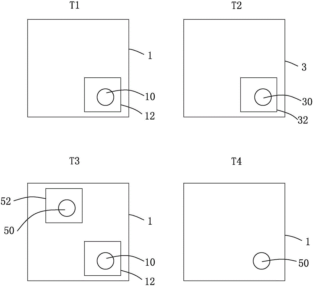
[0072] The utility model (invention) will be further described below in conjunction with the accompanying drawings and embodiments. see figure 1 , is a repair principle diagram of the three-dimensional fine vessel reconstruction method provided by the present invention. The technical problem to be solved in the present invention is to repair the three-dimensional fine blood vessel image with missing parts, which is denoted as image to be repaired 1 . The present invention also provides a reference image 3 for repairing the image 1 to be repaired. Specifically, on the premise of not destroying the blood vessel sample, in order to obtain the microscopic multi-scale image data of the blood vessel sample, a blood vessel sample is serially sliced, and the image to be repaired is obtained by scanning with a high-resolution optical and electron microscope 1. Since the higher the resolution, the smaller the range of the sample area that can be scanned, the image data obtained by mic...


PUM


Abstract

The invention discloses a three-dimensional fine blood vessel reconstruction method. The three-dimensional fine blood vessel reconstruction method comprises the following steps of 1, loading a three-dimensional fine blood vessel original image; 2, performing blood vessel property-based image preprocessing; 3, performing blood vessel property-based characteristic matching; and 4, performing image filling and boundary processing. The invention furthermore discloses a three-dimensional fine blood vessel reconstruction system. With the adoption of the three-dimensional fine blood vessel reconstruction method and system provided by the invention, a filling region for repairing a missing part is obtained by mapping a position relationship and calculating an overall matching degree, and the missing part is repaired through the filling region to obtain a complete high-precision image, so that the accuracy of three-dimensional fine blood vessel reconstruction is high.

Description

technical field [0001] The invention relates to the intersection field of digital image processing and medical imaging, in particular to a three-dimensional fine blood vessel reconstruction method and system thereof. Background technique [0002] In the field of biomedicine, different imaging technologies can observe different phenomena and data, and the advantages of different scanning equipment can be considered comprehensively to obtain microscopic image data of real biological soft tissues at multiple scales. A CT image is a scanned image of a certain part of the human body. It can image blood vessels, tumors and other tissues. Although the imaging is complete, its disadvantage is that the quality of details of the scanned data is obviously not as good as that of tissue slices, and the types of tissues that can be distinguished are limited. Using optical and electron microscopes to image tissue slices can obtain fine-scale multi-scale image data with higher resolution an...

Claims


Application Information

Patent Timeline
IPC IPC(8): G06T17/00
CPCG06T17/00G06T2210/41
Inventor 廖胜辉夏佳志郑晚秋梁毅雄邹北骥李芳芳
Owner CENT SOUTH UNIV
Why Eureka
Try Eureka