
Method and system for machine learning based assessment of fractional flow reserve

A deep neural network, object detection technology, applied in image data processing, instrumentation, computing, etc., can solve problems such as robustness of anatomical object detection

Active Publication Date: 2017-02-15
SIEMENS AG
View PDF4 Cites 24 Cited by
  • Summary
  • Abstract
  • Description
  • Claims
  • Application Information

AI Technical Summary

Problems solved by technology

However, anatomical object detection using MSL is not always robust, especially for some challenging detection problems where anatomical objects exhibit large variations in anatomy, shape, or appearance in medical images

Method used


Image

  • Method and system for machine learning based assessment of fractional flow reserve
  • Method and system for machine learning based assessment of fractional flow reserve
  • Method and system for machine learning based assessment of fractional flow reserve

Examples


Embodiment Construction

[0021] The present invention relates to methods and systems for anatomical object detection using edge-space deep neural networks. Embodiments of the invention are described herein to give an intuitive understanding of a machine learning based approach for anatomical object detection. Digital images usually consist of digital representations of one or more objects (or shapes). Digital representations of objects are generally described herein in terms of recognizing and manipulating objects. Such manipulations are intuitive manipulations implemented in the computer system's memory or other circuitry / hardware. Thus, it should be understood that embodiments of the present invention can be implemented within a computer system using data stored within the computer system.

[0022] Marginal Space Learning (MSL) is an efficient discriminative learning framework that can be used for anatomical object detection and tracking in medical images such as but not limited to computed tomogr...


PUM


Abstract

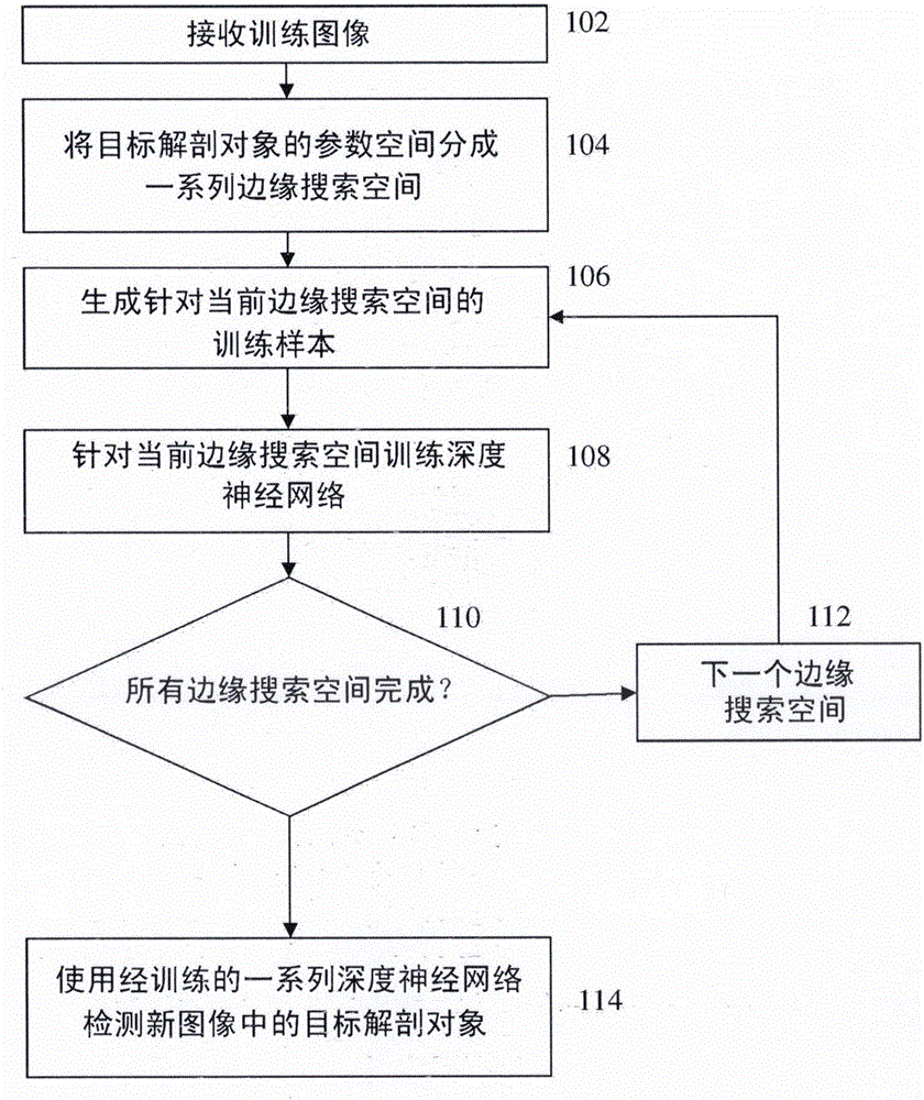
A method and system for anatomical object detection using marginal space deep neural networks is disclosed. The pose parameter space for an anatomical object is divided into a series of marginal search spaces with increasing dimensionality. A respective deep neural network is trained for each of the marginal search spaces, resulting in a series of trained deep neural networks. Each of the trained deep neural networks can evaluate hypotheses in a current parameter space using discriminative classification or a regression function. An anatomical object is detected in a medical image by sequentially applying the series of trained deep neural networks to the medical image.

Description

[0001] This application claims the benefit of U.S. Provisional Application No. 62 / 148,273, filed April 16, 2015, and U.S. Provisional Application No. 62 / 121,782, filed February 27, 2015, and filed October 16, 2014 Continuation-in-Part of filed U.S. Application No. 14 / 516,163 claiming the benefit of U.S. Provisional Application No. 61 / 891,920 filed October 17, 2013, the disclosures of which are incorporated by reference This article. technical field [0002] The present invention relates to anatomical object detection in medical image data, and more particularly, to anatomical object detection in medical image data using deep neural networks. Background technique [0003] Fast and robust anatomical object detection is a fundamental task in medical image analysis that underpins the entire clinical imaging workflow from diagnosis, patient stratification, treatment planning, intervention, and follow-up. Automatic detection of anatomical objects is a prerequisite for many medica...

Claims


Application Information

Patent Timeline
IPC IPC(8): G06T7/77
CPCG06T7/0012G06T2207/20081G06T2207/20084G06T2207/30004G06T7/73G06T2207/10088G06T2207/20021G06T2207/20076G06T2207/30048
Inventor D·科马尼丘B·乔治斯库刘大元H·阮V·K·辛格郑冶枫
Owner SIEMENS AG
Learn More