Method for classifying and identifying visible components of microscopic excrement examination images based on machine vision

A technology of machine vision and classification recognition, which is applied in the field of image processing, can solve the problems of the accuracy and speed of classification and recognition of formed components, lack of objective standards, heavy inspection workload, etc. The effect of fast speed and avoiding boundary leakage

Active Publication Date: 2017-05-17
SICHUAN ORIENTER BIOLOGICAL TECH
View PDF6 Cites 22 Cited by
  • Summary
  • Abstract
  • Description
  • Claims
  • Application Information

AI Technical Summary

Benefits of technology

The technology described in this patented describes an improved way to recognize and sort small parts from large ones called stools during examinations or procedures like xray imagery analysis (XI). By performing these techniques at different stages of the procedure, patient care may become quicker and more accurate than previously possible. Additionally, there have also been developed methods to improve the efficiency of identifying specific areas within each part without relying solely upon visual inspection alone. Overall, this innovative approach improves overall performance and convenience for clinicians while reducing their workload.

Problems solved by technology

In summary, current methods used for identifying stools during medical imaging can lead to errors or delays caused by subjectivity issues such as subjectiveness and lack of standardization across multiple facilities. Additionally, conventional automated systems often require significant effort and resources even if they were able to accurately detect all possible variations within specific areas (regions) without requiring experts manually reviewed data.

Method used


Image

  • Method for classifying and identifying visible components of microscopic excrement examination images based on machine vision
  • Method for classifying and identifying visible components of microscopic excrement examination images based on machine vision
  • Method for classifying and identifying visible components of microscopic excrement examination images based on machine vision

Examples


Embodiment 1

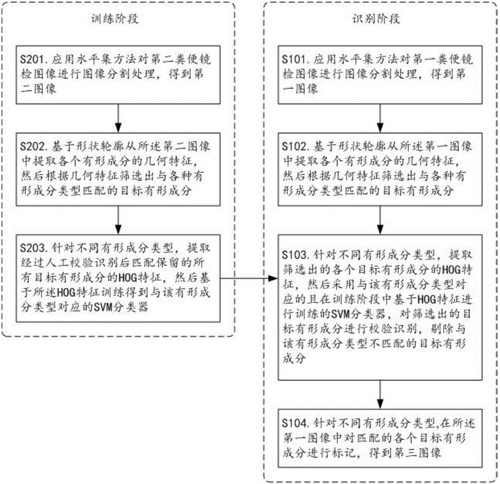
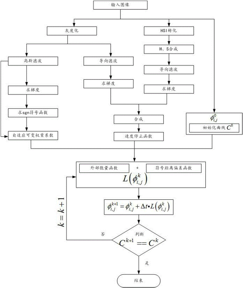
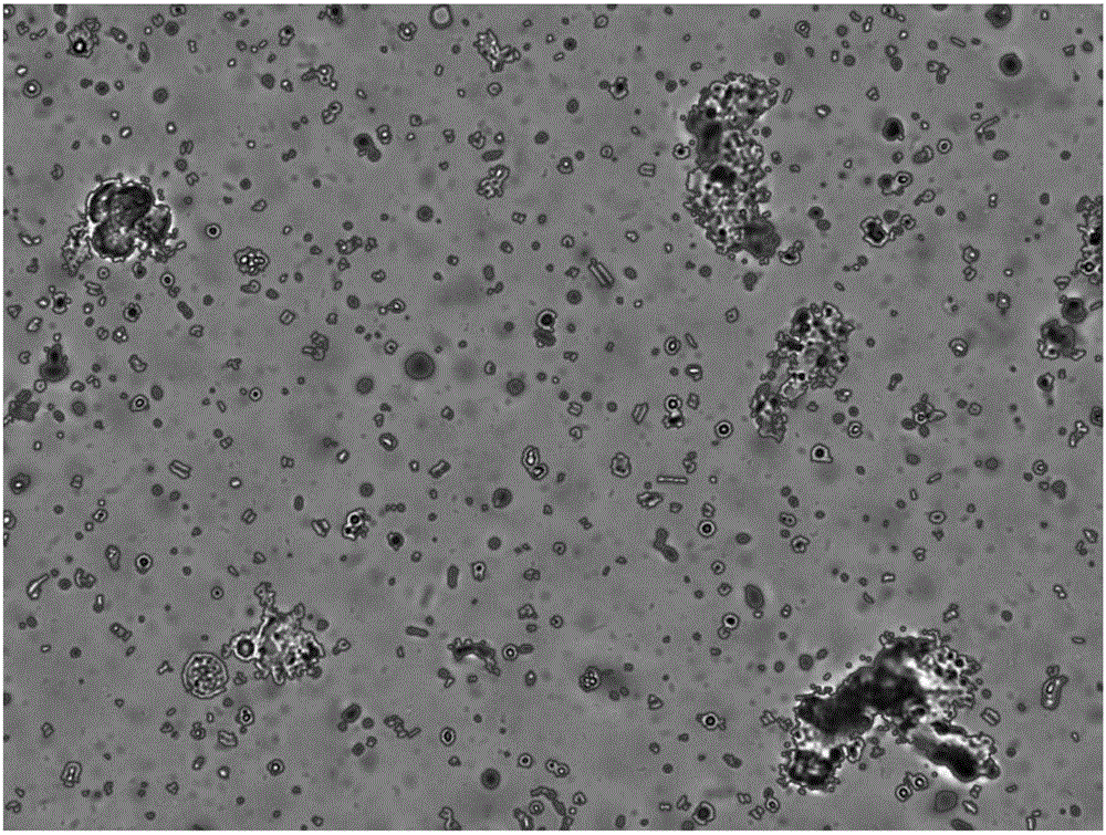
[0043] figure 1 It shows a schematic flowchart of the machine vision-based classification and recognition method for formed components in feces microscopy images provided by the present invention, figure 2 It shows a schematic flow chart of applying the level set method for image segmentation processing provided by the present invention, image 3 It shows an example diagram of an image containing the contour of a formed component shape obtained after applying the level set method for image segmentation processing provided by the present invention, Figure 4 It shows an example diagram of an image containing the classification mark of formed components obtained after classification and identification provided by the present invention. The machine vision-based method for classifying and identifying formed components in feces microscopy images provided in this embodiment includes a training phase and a recognition phase.

[0044] S101. Applying the level set method to perform ...


PUM


Abstract

The invention relates to the technical field of image processing and discloses a method for classifying and identifying visible components of microscopic excrement examination images based on machine vision. Firstly, a level set method is applied to conduct image segmentation on microscopic excrement examination images, then primary classification based on morphological characteristics and verifying identification based on an HOG + VSM are sequentially performed according to shapes and outlines of the visible components obtained through segmentation, and finally high-precision visible component classification and identification results are rapidly obtained for timely consulting of patients. In addition, the method further has the advantages of being high image segmentation precision, high in processing speed, good in user experience and the like and is convenient to apply actually and popularize.

Description


Claims


Application Information

Patent Timeline
Owner SICHUAN ORIENTER BIOLOGICAL TECH
Why Eureka
Try Eureka