Radiation Imaging Apparatus, And Insertion State Determination Method

A technology for medical images and camera devices, applied in medical science, radiation beam steering devices, mammography, etc., can solve the problems of inappropriate, unable to fully insert the breast, difficult to take pictures, etc.

Inactive Publication Date: 2017-09-26
CANON KK
View PDF10 Cites 0 Cited by
  • Summary
  • Abstract
  • Description
  • Claims
  • Application Information

AI Technical Summary

Problems solved by technology

In the upright type breast CBCT, unlike the prone position type, it is impossible to insert the breast into the insertion part by tilting the subject's body using the subject's body weight, making it impossible to fully insert the breast in some cases (i.e., insertion state is inappropriate), and the dead zone changes depending on the insertion state
Variations in blind spots make it difficult to capture desired areas
Another issue is that the blind zone varies more in erect breast CBCT than in mammograms taken with the breast immobilized

Method used


Image

  • Radiation Imaging Apparatus, And Insertion State Determination Method
  • Radiation Imaging Apparatus, And Insertion State Determination Method
  • Radiation Imaging Apparatus, And Insertion State Determination Method

Examples


no. 1 example

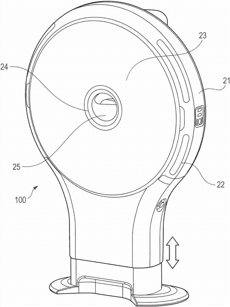
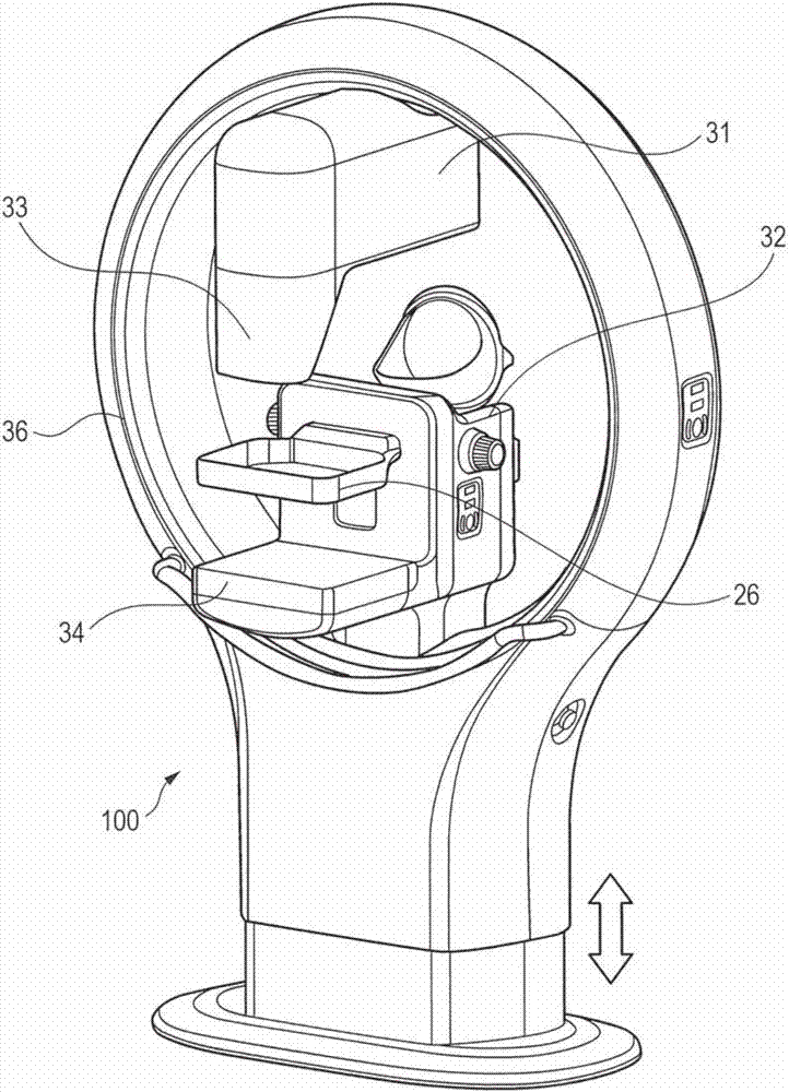
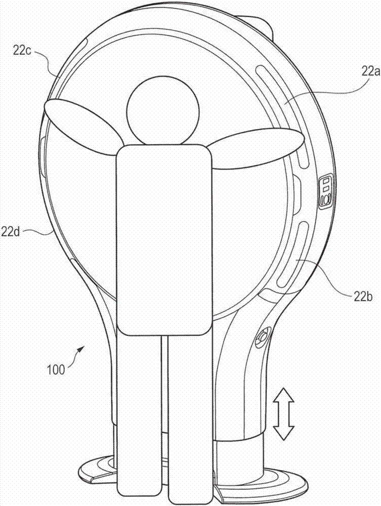
[0053] Figure 7Aa and Figure 7Ab are diagrams each illustrating an example of the insertion state of the breast in the first embodiment. The radiation imaging system (medical imaging system) 100 appropriately manages blind spots in the C area and the C' area (outer upper area), which are areas prone to breast cancer. Except for the case where a radiographic image of the whole body of the subject P is captured by a CT apparatus, it is necessary to properly manage blind spots. In this case, simply put, proper management of the blind zone means setting the blind zone in areas C (outer upper area) and D (outer lower area) of the breast smaller than in area A (inner upper area) of the breast and the dead zone in zone B (inner lower zone).

[0054] Figure 7Aa It is a diagram illustrating a state in which the left breast is inserted into the imaging region of the upright breast CBCT as viewed from the leg side of the subject P. A rotatable frame 36 provided in this part of th...

no. 2 example

[0098] In the radiation imaging system (medical image imaging system) 100 of the first embodiment, the determination unit 104 is based on at least one of the length, area, volume, shape, and pressure of the inserted breast inserted from the insertion portion into the imaging region of the medical image. Or, to determine the insertion status of the breast. On the other hand, in the radiographic imaging system (medical image imaging system) 100 of the second embodiment, the determination unit 104 determines based on at least one of the length, area, volume, shape, and pressure of the non-inserted breast not inserted into the imaging area. Or, to determine the insertion status of the breast. Note that descriptions of components, functions, and operations of the second embodiment similar to the above embodiment are omitted, and differences between the second embodiment and the above-described embodiment are mainly described below.

[0099] Figure 8 is a diagram illustrating an ...


PUM


Abstract

The invention provides a radiation imaging apparatus, and an insertion state determination method. Provided is a medical image imaging apparatus that images a medical image of a breast, including a determination unit configured to determine an insertion state of the breast based on a form of the breast inserted into an imaging area for the medical image from an insertion portion.

Description

technical field [0001] The present invention relates to a radiographic imaging device for capturing radiographic images of breasts, and an insertion state determination method. Background technique [0002] A mammogram apparatus is generally used as an X-ray examination apparatus for breast cancer. However, it is known that in the case of performing mammography of a dense breast (breast with many breasts), lesion parts and breast structures overlap each other, which leads to deterioration in sensitivity and specificity of lesion detection. As techniques for compensating for the disadvantages of mammography, tomosynthesis and CBCT (Cone beam Computed Tomography) apparatuses dedicated to mammography have attracted attention. These devices have a feature of providing a 3D image of the breast, enabling discrete observation of a lesion portion and a breast structure. [0003] In the case of imaging breasts with a CBCT apparatus dedicated to imaging breasts, it is necessary to m...

Claims


Application Information

Patent Timeline
Patent Type & Authority Applications(China)
IPC IPC(8): A61B6/00A61B6/03
CPCA61B6/032A61B6/44A61B6/502A61B5/0091A61B5/1072A61B5/1073A61B5/1077A61B5/1079A61B6/037A61B6/0414A61B6/0492A61B6/08A61B6/4085A61B6/46A61B6/462A61B6/463A61B6/544A61B6/589A61B8/0825A61B8/403A61B2562/0247
Inventor 辻井修
Owner CANON KK
Why Eureka
Try Eureka