Medical image segmentation method and device

A medical image and image technology, applied in the field of image processing, can solve the problems of poor segmentation effect and low efficiency, and achieve the effect of fast speed, high time efficiency and strong noise resistance

Inactive Publication Date: 2017-10-24
BEIJING INSTITUTE OF TECHNOLOGYGY
View PDF4 Cites 9 Cited by
  • Summary
  • Abstract
  • Description
  • Claims
  • Application Information

AI Technical Summary

Problems solved by technology

[0005] At present, most of these algorithms have certain shortcomings, such as poor segmentation effect and l

Method used


Image

  • Medical image segmentation method and device
  • Medical image segmentation method and device
  • Medical image segmentation method and device

Examples


Embodiment Construction

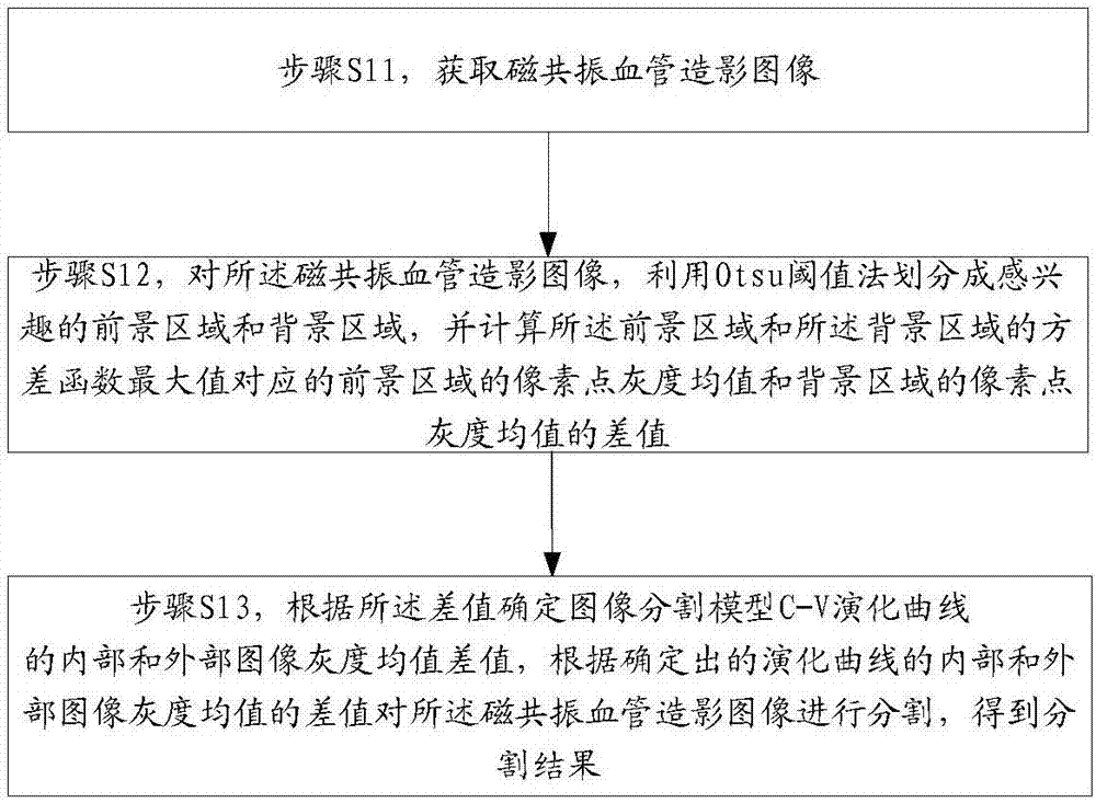
[0021] One image segmentation algorithm is the Otsu threshold method, which was proposed by the Japanese scholar Otsu in 1979, also known as the Otsu method or the maximum inter-class variance method. The Otsu threshold method divides the image into two parts, the background and the target (that is, the region of interest) according to the grayscale characteristics of the image. The greater the inter-class variance between the background and the target, the greater the difference between the two parts of the image. When the image is segmented, part of the target is misclassified as the background or part of the background is misclassified as the target, which will cause the difference between the two parts to change. Small. Therefore, ensuring the maximum variance between classes in image segmentation means the minimum probability of misclassification. The Otsu threshold method has high operation efficiency and fast speed, but the Otsu algorithm itself is sensitive to noise a...


PUM


Abstract

The invention discloses a medical image segmentation method and a device, wherein the method comprises: step S11) obtaining magnetic resonance angiography image; step S12) for the magnetic resonance angiography image, using the Otsu threshold method to divide it into an interested foreground area and a background area; and calculating difference value between the pixel mean gray value of the foreground area and the pixel mean gray value of the background area corresponding to the maximum of the variance function of the foreground area and the background area; and Step S13): determining the difference value between the gray mean values of the internal images and external images of the evolution curve of the image segmentation model C-V according to the difference value; and segmenting the magnetic resonance angiography image according to the determined difference value between the gray mean values of the internal images and external images of the evolution curve and obtaining a segmenting result. The medical image segmentation device includes an image obtaining module, a difference value determining module, and a segmenting module. The medical image segmentation method and the device of the present disclosure improve the segmenting effect and the processing speed, meeting the requirements.

Description

technical field [0001] The invention relates to the technical field of image processing, in particular to a medical image segmentation method and device. Background technique [0002] Medical images are different from ordinary images. The blood vessels themselves have a complex system structure and are easily affected by noise. In addition, due to the differences in human body structures, even for the same organ tissue, the corresponding images may have different There is a large difference, which makes medical image processing more difficult than general image processing. [0003] Image segmentation is the process of dividing an image into several specific regions with unique properties and extracting objects of interest. It is a key step from image processing to image analysis. [0004] Classical image segmentation algorithms can be roughly divided into threshold segmentation method, region growing method and edge detection method, while image segmentation algorithm base...

Claims


Application Information

Patent Timeline
IPC IPC(8): G06T7/11G06T7/136G06T7/194
CPCG06T7/11G06T7/136G06T7/194G06T2207/10088G06T2207/30101
Inventor 马锐刘月马科薛静锋菅泽峰高浩然
Owner BEIJING INSTITUTE OF TECHNOLOGYGY
Why Eureka
Try Eureka