Methods of Prostate Segmentation in Medical Images

A medical image, prostate technology, applied in the field of medical images, to achieve the effect of reliable boundary information

Active Publication Date: 2021-01-29
SHANGHAI UNITED IMAGING HEALTHCARE
View PDF11 Cites 0 Cited by
  • Summary
  • Abstract
  • Description
  • Claims
  • Application Information

AI Technical Summary

Problems solved by technology

[0003] The task of prostate segmentation is particularly challenging

Method used


Image

  • Methods of Prostate Segmentation in Medical Images
  • Methods of Prostate Segmentation in Medical Images
  • Methods of Prostate Segmentation in Medical Images

Examples


Embodiment Construction

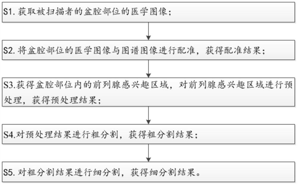
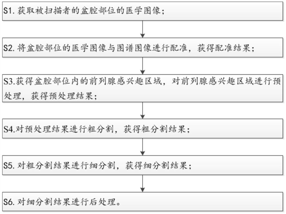
[0049] see figure 1 , 3-5, the prostate segmentation method in a kind of medical image of the embodiment of the present invention, comprises the following steps:

[0050] S1. Obtain a medical image of the pelvic region of the scanned person;

[0051] S2. Register the medical image of the pelvic region with the atlas image to obtain a registration result;

[0052] S3. Obtain the prostate region of interest in the pelvic cavity, perform preprocessing on the prostate region of interest, and obtain a preprocessing result;

[0053] S4. Roughly segment the preprocessing result to obtain a rough segment result;

[0054] S5. Perform fine segmentation on the rough segmentation result to obtain the fine segmentation result.

[0055] Obtaining the medical image of the pelvic region of the scanned person in the embodiment of the present invention includes the following steps:

[0056] S11. Input one of the CT image, MR image or DR image of the pelvic region of the scanned person;

[...


PUM


Abstract

The invention discloses a prostate segmentation method in medical images, comprising the following steps: S1. Obtaining a medical image of the pelvic region of a scanned person; S2. Registering the medical image of the pelvic region with an atlas image to obtain a registration result ; S3. Obtain the prostate region of interest in the pelvic cavity, preprocess the prostate region of interest, and obtain the preprocessing result; S4. Perform rough segmentation on the preprocessing result, and obtain the rough segmentation result; S5. Fine-tune the rough segmentation result Segmentation obtains fine segmentation results, and the method of the present invention can realize automatic segmentation of the prostate.

Description

【Technical field】 [0001] The invention relates to the field of medical images, in particular to the segmentation of the prostate in the medical images. 【Background technique】 [0002] An essential prerequisite for the successful treatment of cancer with computer-assisted radiation therapy is to confirm the location and size of the tumor while preserving critical organs around the lesion. Therefore, accurate and efficient extraction of key organ contours and calculation of gross tumor volume (GTV, gross tumor volume) are key steps in adaptive radiation therapy and surgical navigation, and have very important research significance. When lesions occur around the genitals, the genitals must be drawn separately for protection to avoid radiation exposure. Therefore, when tumors appear in the male pelvic cavity, the prostate is the first organ to avoid urgently. Moreover, prostate cancer is the most common malignant tumor of the male reproductive system, and its morbidity and mor...

Claims


Application Information

Patent Timeline
Patent Type & Authority Patents(China)
IPC IPC(8): G06T7/10G06T7/136G06T7/30
Inventor 马金凤韩妙飞李强
Owner SHANGHAI UNITED IMAGING HEALTHCARE
Why Eureka
Try Eureka