Automatic Segmentation Method of Eye Cup in Fundus Image Based on Improved PDE Image Inpainting

A fundus image, automatic segmentation technology, applied in the field of medical image processing, can solve the problems of the segmentation results remaining in the preliminary stage, the segmentation results are not accurate, and the edge of the optic cup is not clear, so as to reduce manual intervention, ensure accuracy, and ensure The effect of split speed

Inactive Publication Date: 2018-10-12
BEIJING UNIV OF TECH
View PDF2 Cites 0 Cited by
  • Summary
  • Abstract
  • Description
  • Claims
  • Application Information

AI Technical Summary

Problems solved by technology

[0004] However, the current methods for automatic segmentation of the optic cup are still very limited. In the process of segmentation of the optic cup, the current method is easily affected by the unclear edge of the optic cup caused by blood vessels, resulting in inaccurate segmentation results. Therefore, the existing The segmentation results of the method are still in the preliminary stage

Method used


Image

  • Automatic Segmentation Method of Eye Cup in Fundus Image Based on Improved PDE Image Inpainting
  • Automatic Segmentation Method of Eye Cup in Fundus Image Based on Improved PDE Image Inpainting
  • Automatic Segmentation Method of Eye Cup in Fundus Image Based on Improved PDE Image Inpainting

Examples


Embodiment Construction

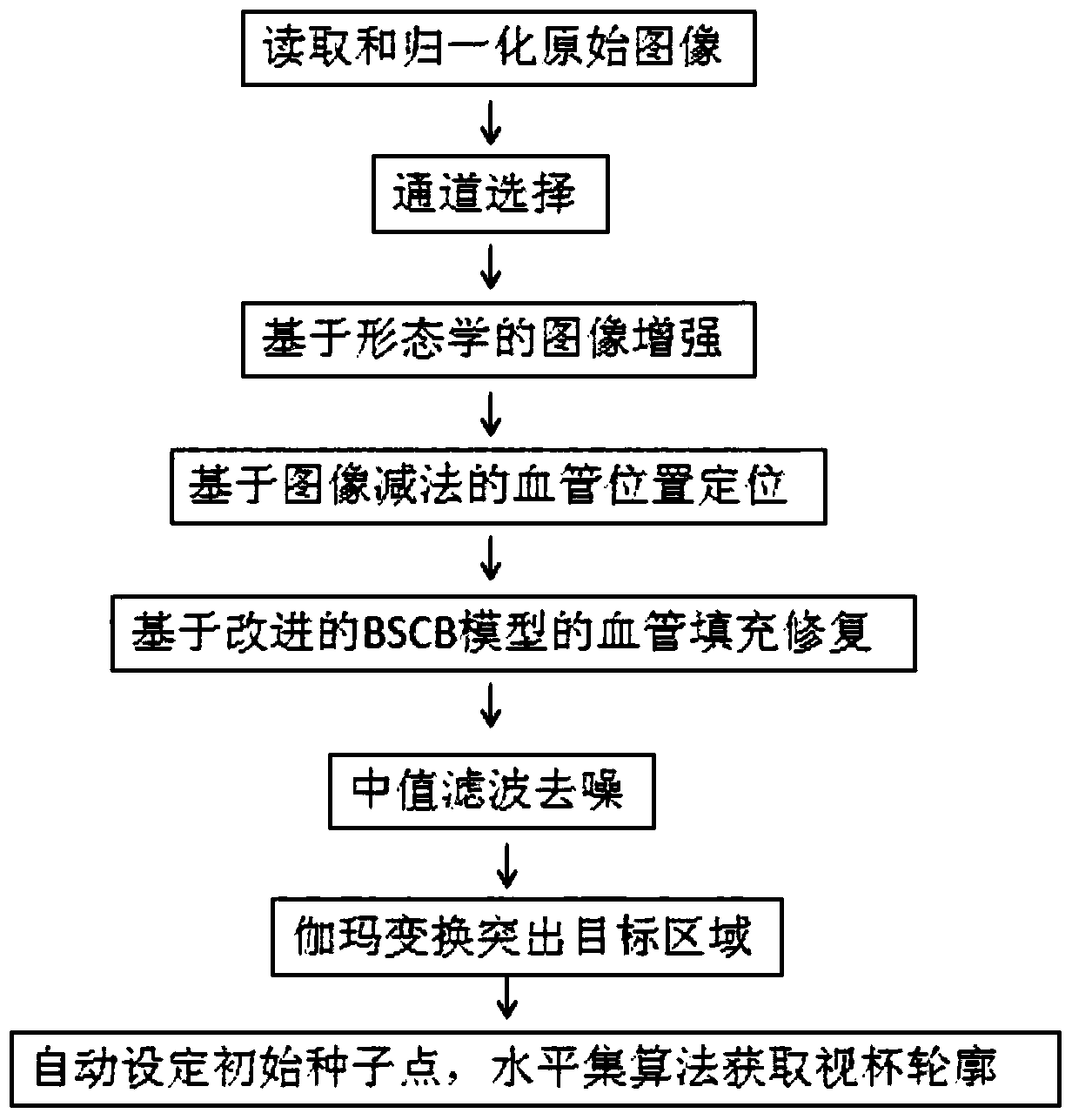
[0029] Step 1, read the original image, and normalize the image size to 1440*960 pixels, such as figure 2 .

[0030] Step 2, perform channel selection on the original image, and select the most prominent green channel image U in the target area G Segmentation of the optic cup, such as image 3 .

[0031] Step 3, the image is enhanced through the principle of morphology. This method uses the image top hat and bottom hat transformation to enhance the image:

[0032]

[0033] B hat (U G )=(U G ·SE)-U G

[0034] u CE =(U G +T hat (U G ))-B hat (U G )

[0035] In the formula: symbol Indicates that the image is opened, and the symbol "·" indicates that the image is closed. SE is a structural element, and a circular structural element of 5*5 is taken. T hat (U G ) is the image after top-hat transformation, B hat (U G ) is the image after bottom hat transformation, U CE is the image after contrast enhancement, and the image after image enhancement is as foll...


PUM


Abstract

The invention discloses an eye fundus image optical cup automatic segmentation method based on improved PDE image repairing. The method is carried out with following steps: reading and normalizing an original image; selecting channels for the original image; enhancing the image based on a morphological principle; positioning blood vessels based on an image subtraction; filling and repairing blood vessel areas by the use of an improved BSCB model; performing median filtering and noise reduction for the image and highlighting object areas through gamma transformation; and preliminarily obtaining an optical cup contour through a level set algorithm under a condition that the mass center of the brightest pixel point set on the image serves as an initial seed point. According to the invention, the image in which the blood vessels are filled and repaired eliminate the influence on optical cup segmentation by the blood vessel areas to a certain extent; the accuracy of optical disk segmentation is improved; the seed point is automatically selected as the contour of an optical cup is obtained through the level set algorithm; and manual intervention in a conventional semi-automatic method is reduced.

Description

technical field [0001] The invention belongs to the field of medical image processing, relates to an improved partial differential equation (PDE)-based BSCB image repair model, and realizes the automatic segmentation of the optic cup of the fundus image. Background technique [0002] Fundus and optic nerve tissue lesions caused by various reasons are called fundus optic nerve diseases (such as glaucoma, diabetic retinopathy, etc.), and these diseases all have the risk of blindness. The incidence of such diseases is very high worldwide, and it is showing an upward trend, and the change of the cup size in the fundus image has important guiding significance for the early diagnosis and treatment of glaucoma and other lesions. Segmentation is a key technique for computer-aided diagnosis of eye-related lesions. [0003] At present, there are many researches on the automatic segmentation of the optic cup in fundus images. These methods are generally divided into three types: meth...

Claims


Application Information

Patent Timeline
Patent Type & Authority Patents(China)
IPC IPC(8): G06T7/11
CPCG06T7/12G06T2207/10004G06T2207/20032G06T2207/20192G06T2207/30041
Inventor 杨春兰段彦华刘冰
Owner BEIJING UNIV OF TECH
Why Eureka
Try Eureka