
A Sequential Pulmonary Nodule Image Segmentation Method Based on Superpixel and Density Clustering

A sequence image and density clustering technology, which is applied in image analysis, image enhancement, image data processing, etc., can solve the problem of large differences in gray value, the inability to efficiently segment the sequence of pulmonary nodule images, and not reduce the accuracy of segmentation And other issues

Active Publication Date: 2019-11-08
TAIYUAN UNIV OF TECH
View PDF2 Cites 0 Cited by
  • Summary
  • Abstract
  • Description
  • Claims
  • Application Information

AI Technical Summary

Problems solved by technology

In the previous research on sequential pulmonary nodule image segmentation algorithms, the segmentation of solid nodules is very effective, but the following problems arise: without reducing the segmentation accuracy, the sequential pulmonary nodule images cannot be efficiently segmented ; There is a large difference in the gray value of the cavity inside the cavitary nodule and the surrounding area, and it is easy to regard the cavity as a part of the lung parenchyma, resulting in incomplete segmentation of the nodule area; the gray value of the vascular adhesion type nodule and the blood vessel The gray value is very close, it is easy to keep the blood vessel as part of the nodule, and cannot effectively separate the blood vessel from the nodule; there is a lot of noise, which leads to the segmentation of the pulmonary nodule and the noise and various tissue structures Therefore, a method that can not only achieve accurate segmentation of sequential pulmonary nodule images, but also meet the clinical requirements in terms of speed has important reference value for assisting physicians to make reliable diagnostic decisions

Method used


Image

  • A Sequential Pulmonary Nodule Image Segmentation Method Based on Superpixel and Density Clustering
  • A Sequential Pulmonary Nodule Image Segmentation Method Based on Superpixel and Density Clustering
  • A Sequential Pulmonary Nodule Image Segmentation Method Based on Superpixel and Density Clustering

Examples


Embodiment Construction

[0078] The present invention will be described in detail below in conjunction with specific examples.

[0079] The realization process of the inventive method is as follows:

[0080] A method for sequential pulmonary nodule image segmentation, comprising the following steps:

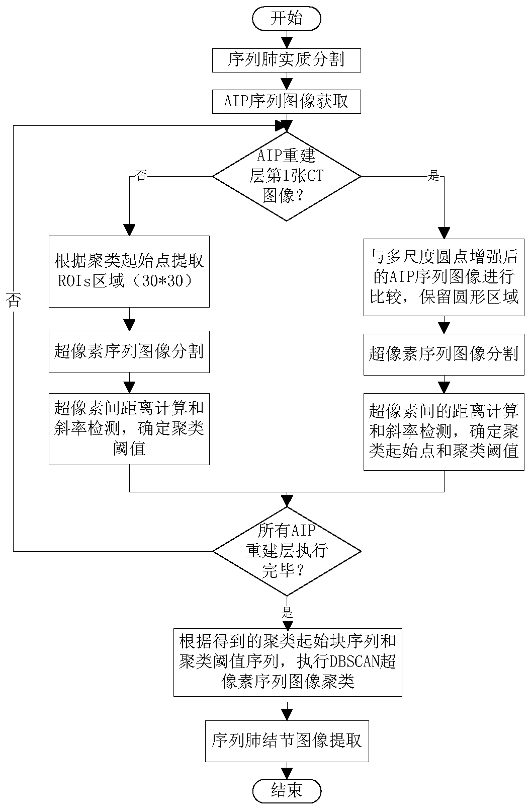
[0081] refer to figure 1 , step A, using CT image three-dimensional feature average projection density (AIP) combined with multi-scale dot enhancement for preprocessing;

[0082] refer to Figure 13 , Figure 14 , Figure 15 In columns (c) and (d), in step B, an improved superpixel segmentation algorithm suitable for lung images is adopted according to the circular and area features of lung nodules in lung images, that is, based on Hexagonal clustering and morphologically optimized sequential linear iterative clustering (HMSLIC) for over-segmentation of lung CT sequence images;

[0083] refer to figure 2 , image 3 , Figure 4 , Figure 5 , Image 6 , Figure 7 , Figure 8 , Figure 9 , ...


PUM


Abstract

The invention discloses a sequential pulmonary nodule image segmentation method based on superpixels and density clustering, which comprises the following steps: using CT image three-dimensional feature average projection density (AIP) combined with multi-scale dot enhancement for preprocessing; using hexagonal The superpixel sequential image segmentation algorithm (HMSLIC) with shape clustering and morphological optimization (HMSLIC) is used to over-segment the image; in the calculation of the distance between superpixel blocks, an adaptive weight coefficient is constructed to realize the precise positioning of pulmonary nodules and obtain subsequent clustering At the same time, by fitting the distance and detecting the change of the slope, the subsequent accurate clustering threshold is obtained; the fast DBSCAN superpixel optimized by only clustering the lung nodules and adaptive threshold is adopted Sequential image clustering algorithm to obtain sequential pulmonary nodule masks, and finally obtain sequential pulmonary nodule images of lung CT; it can more quickly, completely and accurately segment various types of sequential pulmonary nodule images for subsequent processing and diagnostic analysis.

Description

technical field [0001] The invention relates to the segmentation of pulmonary nodule sequence images, in particular to a sequential pulmonary nodule image segmentation method based on superpixels and density clustering. Background technique [0002] Fast and accurate segmentation of pulmonary nodule sequence images is the basis for subsequent processing and diagnostic analysis. In the previous research on sequential pulmonary nodule image segmentation algorithms, the segmentation of solid nodules is very effective, but the following problems arise: without reducing the segmentation accuracy, the sequential pulmonary nodule images cannot be efficiently segmented ; There is a large difference in the gray value of the cavity inside the cavitary nodule and the surrounding area, and it is easy to regard the cavity as a part of the lung parenchyma, resulting in incomplete segmentation of the nodule area; the gray value of the vascular adhesion type nodule and the blood vessel The...

Claims


Application Information

Patent Timeline
Patent Type & Authority Patents(China)
IPC IPC(8): G06T7/174G06T7/187G06T7/64G06K9/62
CPCG06T7/174G06T7/187G06T7/64G06T2207/30064G06T2207/20021G06T2207/10081G06F18/23
Inventor 强彦张伟赵涓涓宋晓涛强梓林
Owner TAIYUAN UNIV OF TECH
Learn More