Ultrasonic low-frequency transcranial volume super-resolution radiographic imaging method and system

A super-resolution and contrast-enhanced imaging technology, applied in the fields of ultrasound/sonic/infrasonic Permian technology, ultrasound/sonic/infrasonic image/data processing, ultrasound/sonic/infrasonic diagnosis, etc. There are problems such as clear super-resolution imaging technology and insufficient resolution

Inactive Publication Date: 2018-07-27
XI AN JIAOTONG UNIV
View PDF11 Cites 16 Cited by
  • Summary
  • Abstract
  • Description
  • Claims
  • Application Information

AI Technical Summary

Problems solved by technology

[0004] Three-dimensional ultrasound imaging is also called ultrasound volume imaging. According to different acquisition methods of ultrasound three-dimensional volume data, it can be divided into two types: one is static imaging, that is, the two-dimensional signal of the space section is obtained by scanning the mechanically controlled probe, and then the two-dimensional The signal is reconstructed in space to obtain a three-dimensional image. This method has high resolution, but to a certain extent, time information is lost, and an accurate positioning system is required; the second is dynamic imaging, that is, volume imaging technology, which uses area array volume probes Emitting spatial ultrasonic beams to directly obtain 3D ultrasonic volume data, not only has all the functions of a 2D color Doppler ultrasound transducer, but als

Method used


Image

  • Ultrasonic low-frequency transcranial volume super-resolution radiographic imaging method and system
  • Ultrasonic low-frequency transcranial volume super-resolution radiographic imaging method and system
  • Ultrasonic low-frequency transcranial volume super-resolution radiographic imaging method and system

Examples


Embodiment Construction

[0084] The present invention will be further described in detail below in conjunction with the accompanying drawings and embodiments.

[0085] The present invention proposes a transducer array design for transcranial dedicated small area volume imaging, and a transcranial volume super-resolution imaging method based on the transducer array.

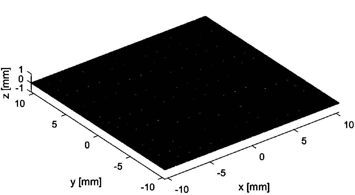
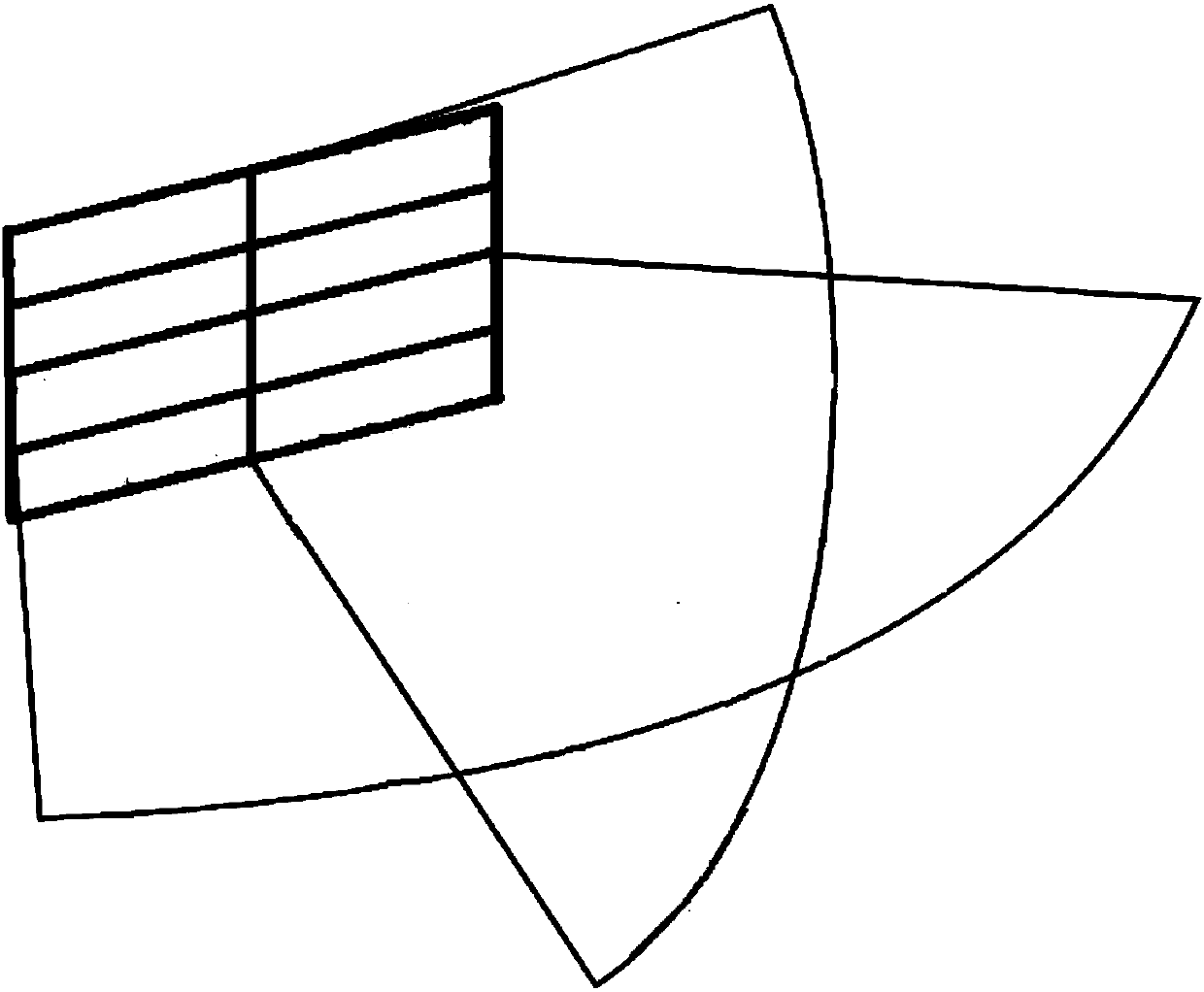
[0086] see figure 1 , the transcranial dedicated small-area volume imaging transducer adopts the ultrasonic small-area two-dimensional planar transducer array design: by N 1 ×N 2 The size of each array element is l×l, the distance between the center of the array element is d, and the distance between the focal point and the geometric center of the surface array is F. see figure 2 , the volume imaging transducer is a transcranial special low-frequency ultrasonic volume transducer, which adopts a two-dimensional array transducer, can scan in two directions when using the transducer, and can collect a scan within a certain range All the...


PUM

PropertyMeasurementUnit
Center frequencyaaaaaaaaaa

Abstract

The invention provides an ultrasonic low-frequency transcranial volume super-resolution radiographic imaging method and system. The volume imaging technique is applied to the field of transcranial angiography; a two-dimensional plane transducer emits ultrasonic plane wave into the skull; scanning is performed in two directions perpendicular to each other; scanned images are subjected to transcranial vascular small-region three-dimensional super-resolution imaging by means of a voxel nearest neighbors process and a multi-objective tracking algorithm based on Markov chain Monte Carlo so as to obtain real-time high-resolution three-dimensional imaging for cranial internal structure.

Description

technical field [0001] The invention belongs to the technical field of ultrasonic detection and ultrasonic imaging, and in particular relates to an ultrasonic two-dimensional planar transducer array and a transcranial low-frequency super-resolution volume three-dimensional imaging method. Background technique [0002] At present, the main means of ultrasonic monitoring in the skull is the transcranial Doppler technology, which uses the frequency shift information of the echo signal to deduce the relative motion between the ultrasonic source and the scattering or reflecting target, and is used to detect the main components of the brain base. Arterial hemodynamics and blood flow physiological parameters. However, transcranial Doppler technology can only display two-dimensional information of vascular blood flow. [0003] Compared with traditional transcranial 2D imaging technology, transcranial 3D imaging technology has many advantages: firstly, 3D imaging is more intuitive a...

Claims


Application Information

Patent Timeline
IPC IPC(8): A61B8/08A61B8/14
CPCA61B8/0891A61B8/14A61B8/4483A61B8/466A61B8/5269
Inventor 万明习柏晨张馨予乔晓阳纪美伶
Owner XI AN JIAOTONG UNIV
Why Eureka
Try Eureka