Method for identifying unconventional cells in pathological section

A technology of pathological slices and identification methods, applied in the field of identification of unconventional cells in pathological slices, can solve the problems of increased error rate, heavy work, time-consuming, etc., and achieve effective classification and good test results

Active Publication Date: 2018-07-31
ZHEJIANG UNIV
View PDF7 Cites 34 Cited by
  • Summary
  • Abstract
  • Description
  • Claims
  • Application Information

AI Technical Summary

Problems solved by technology

[0002] Unconventional cells (or cells with abnormal shapes) in traditional pathological slices are screened manually: under a microscope, professional pathologists move the slices, and then scan the en

Method used


Image

  • Method for identifying unconventional cells in pathological section
  • Method for identifying unconventional cells in pathological section
  • Method for identifying unconventional cells in pathological section

Examples


Embodiment Construction

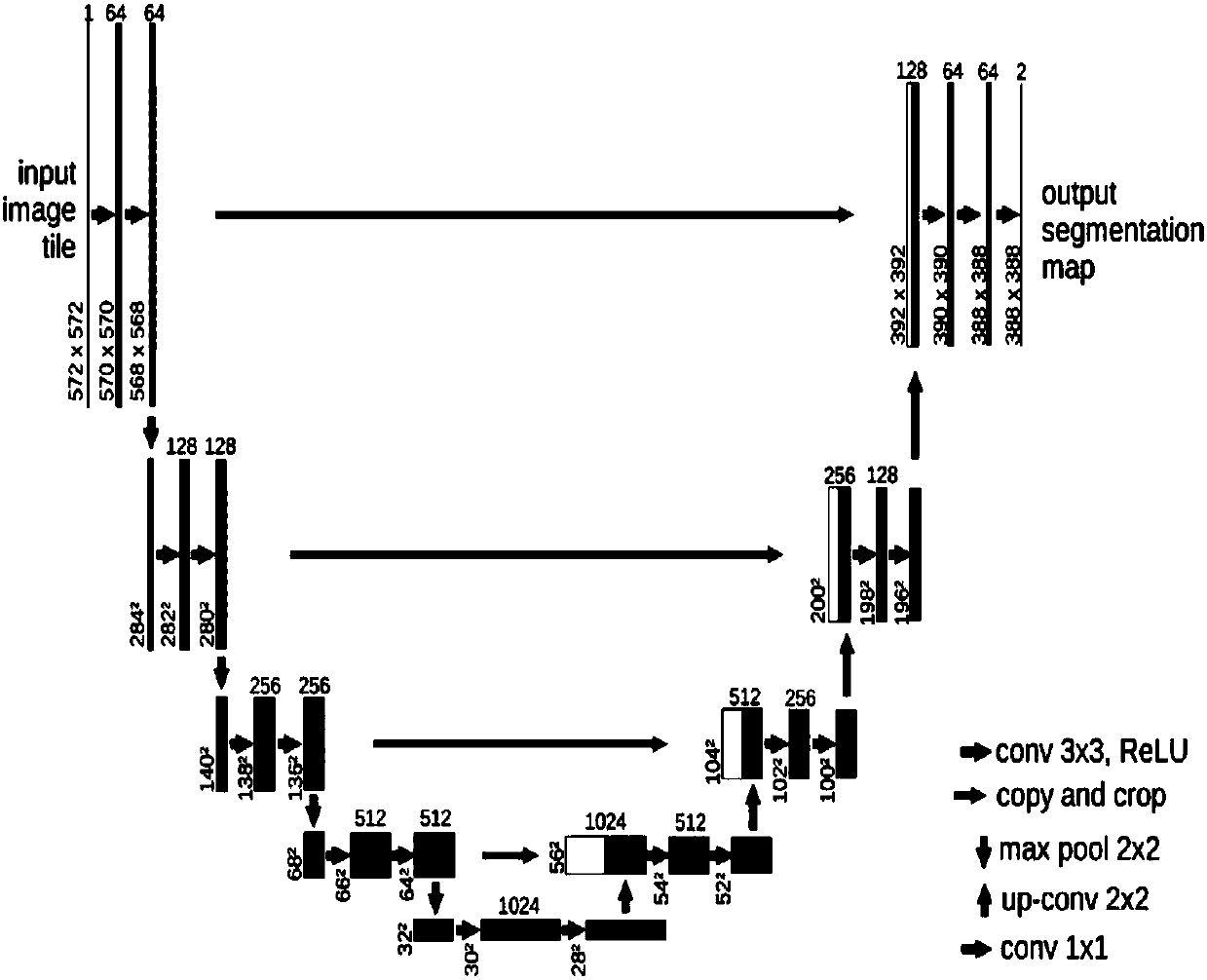
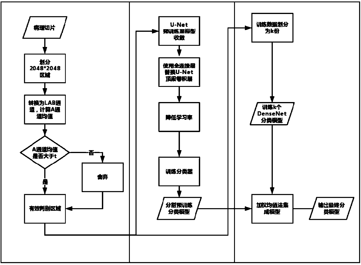
[0068] In order to further understand the present invention, the method for identifying unconventional cells in a pathological section provided by the present invention will be specifically described below in conjunction with specific implementation methods, but the present invention is not limited thereto. The non-essential improvements and adjustments mentioned above still belong to the protection scope of the present invention.

[0069] A method for identifying unconventional cells on pathological slices, the specific steps are:

[0070] 1) Pathological slice preprocessing and valid area discrimination

[0071] In the present invention, the input data is 20x enlarged pathological slices, which are divided into areas with a pixel resolution of 2048*2048 and stored separately.

[0072] Convert the region with a pixel resolution of 2048*2048 above into LAB channels, and use the region where the average value of channel A exceeds the threshold t=132 as an effective discriminat...


PUM


Abstract

The invention discloses a method for identifying unconventional cells in a pathological section, comprising: preprocessing an electronic scanning pathological section to obtain an effective discriminating region, inputting the effective discriminating region into a full convolution network to be pre-trained, then replacing the full convolution network head fine-tuning network with a fully connected layer so that the full convolution network has an ability to extract unconventional cell features so as to determine the locations of unconventional cells and classify the effective discriminatingregions effectively; in combination with the prediction result vote of multiple common classification networks, outputting stable classification results. The identification method of the invention canautomatically discriminate the probability of existence of unconventional cells in each 20* magnified field of view in the pathological section, and outputs the unconventional cells with a probability value over 0.5 as a recognition result, greatly reduce workload of the artificial screening pathological section, and can quickly and accurately screen out the unconventional cells.

Description

technical field [0001] The invention belongs to the field of medical imaging, in particular to a method for identifying unconventional cells in pathological slices. Background technique [0002] Unconventional cells (or cells with abnormal shapes) in traditional pathological slices are screened manually: under a microscope, professional pathologists move the slices, and then scan the entire slice with naked eyes to find whether there are unconventional cells in the entire slice. This work is tedious and time-consuming, and as the reading time increases, the error rate also increases. [0003] With the continuous development of technology, the identification of unconventional cells in pathological slides can be initially screened with the help of computers. [0004] Based on the Convolutional Neural Network (CNN) algorithm, VGGNet, ResNet, DenseNet and other network improvement structures are constantly updated and iterated for computer vision. On natural images, its accurac...

Claims


Application Information

Patent Timeline
IPC IPC(8): G06T7/00G06T7/11G06T3/40G06N3/04
CPCG06T3/4007G06T7/0012G06T7/11G06T2207/20081G06T2207/30024G06N3/045
Inventor 吴健王彦杰王文哲刘雪晨吴边陈为吴福理吴朝晖
Owner ZHEJIANG UNIV
Why Eureka
Try Eureka