Breast mass automatic detection method and system

An automatic detection and mass detection technology, applied in mammography, medical science, image data processing, etc., can solve the problems of delaying the patient's treatment time, difficult to detect tiny calcifications of breast cancer in time, etc., to achieve strong adaptability and accurate detection. Effect

Active Publication Date: 2018-08-31
讯飞医疗科技股份有限公司
View PDF7 Cites 24 Cited by
  • Summary
  • Abstract
  • Description
  • Claims
  • Application Information

AI Technical Summary

Problems solved by technology

Although mammography has many of the above advantages, it still has some unavoidable defects. In particular, only a small part of the information of mammography images can be recognized by human eyes, and it is difficult for experienced doctors to find mammography in time. Tiny calcifications in early-stage breast cancer on X-ray images, delaying treatment for patients

Method used


Image

  • Breast mass automatic detection method and system
  • Breast mass automatic detection method and system
  • Breast mass automatic detection method and system

Examples


Embodiment Construction

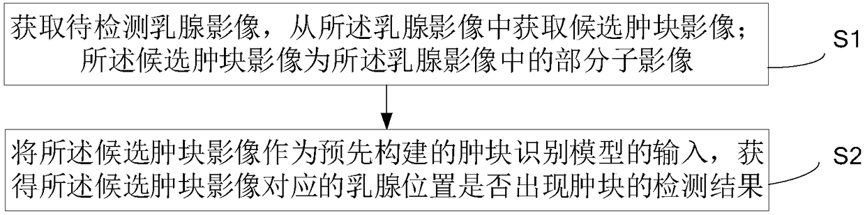
[0023] The specific implementation manners of the present invention will be further described in detail below in conjunction with the accompanying drawings and embodiments. The following examples are used to illustrate the present invention, but are not intended to limit the scope of the present invention.

[0024] With the development of image recognition technology, the application fields of image recognition technology are becoming wider and wider. At present, image recognition technology has been applied in the medical field. However, due to the complexity of breast medical images (images that take into account factors such as shooting quality, patient's physical condition, and shooting technology), the existing manual reading methods, due to the existence of a large amount of image data, not only bring great difficulties to radiologists. Very heavy workload, reading fatigue leads to judgment errors, and the reading results will also be limited by the professional level o...


PUM


Abstract

An embodiment of the invention provides a breast mass automatic detection method and system. The method includes acquiring a to-be-detected breast image and acquiring a mass image from the breast image, wherein the candidate mass image is a part of sub images in the breast image; obtaining a detection result about whether a breast position corresponding to the candidate mass image has a mass or not by taking the candidate mass image as the input of a pre-constructed mass recognition model. The method can implement segmentation of the to-be-detected image more accurately. At the same time, by utilizing a neural network recognition model constructed in advance according to a large amount of sample data for recognizing the image subjected to segmentation, the breast mass is acquired. The method provided by the invention has beneficial effects of good adaptability and accurate detection.

Description

technical field [0001] The invention relates to the technical field of image information processing, and more specifically, to a method and system for automatic detection of breast masses. Background technique [0002] With the continuous improvement of the medical level, people will have various image data to assist in the diagnosis of diseases during the medical treatment process. Relevant statistics indicate that imaging data accounts for 90% of hospital data and is increasing at a rate of 30% per year, while radiologists are only increasing at a rate of about 4%. It can be seen that the review of a large amount of imaging data Bring great workload to the doctor. In addition, if doctors review image data for a long time, they are prone to fatigue, which may lead to possible errors in diagnosis. [0003] Breast lesions are a common disease that threatens women's health. In particular, the incidence of breast cancer has jumped to the top of the incidence of female maligna...

Claims


Application Information

Patent Timeline
Patent Type & Authority Applications(China)
IPC IPC(8): A61B6/00G06T7/10G06T7/44
CPCA61B6/502A61B6/5217G06T7/0012G06T2207/10116G06T2207/20081G06T2207/30068G06T7/10G06T7/44
Inventor 王凤艳殷保才刘聪
Owner 讯飞医疗科技股份有限公司
Why Eureka
Try Eureka