
Method for automatically identifying urinary sediment visible components based on Trimmed SSD

A urinary sediment and tangible technology, which is applied in the field of automatic recognition of the formed components of urine sediment, can solve problems such as not seeing the automatic recognition field

Inactive Publication Date: 2018-10-09
CENT SOUTH UNIV
View PDF2 Cites 12 Cited by
  • Summary
  • Abstract
  • Description
  • Claims
  • Application Information

AI Technical Summary

Problems solved by technology

However, there is no research and attempt in the field of automatic recognition of the formed components of urine sediment.

Method used


Image

  • Method for automatically identifying urinary sediment visible components based on Trimmed SSD
  • Method for automatically identifying urinary sediment visible components based on Trimmed SSD
  • Method for automatically identifying urinary sediment visible components based on Trimmed SSD

Examples


Embodiment Construction

[0064] The present invention will be further described below with reference to the accompanying drawings and examples.

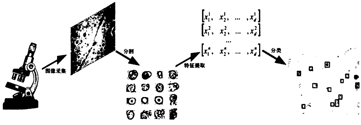
[0065] like figure 1 As shown, existing methods for automated analysis of urine microscopic images employ a traditional multi-stage identification process, including three main stages of segmentation, manual feature extraction, and classifier training. Although there are a large number of algorithms to choose from at each stage, the performance of these algorithms for urine sediment microscopic images is largely determined by the adaptation and tight fit of each stage, the accuracy of target region segmentation and handcrafted features. The effectiveness of the design is especially critical.

[0066] like figure 2 As shown, the present invention regards the identification of formed components of urine sediment as the detection problem of objects, and effectively integrates segmentation, feature extraction and classification into one network by constructin...


PUM


Abstract

The invention discloses a method for automatically identifying urinary sediment visible components based on Trimmed SSD. The method comprises steps that firstly, local detail features and global semantic features are extracted by the feature extraction network composed of the basic convolutional network and the auxiliary convolutional network; secondly, three feature maps are selected by the target identification network as input from different layers of the feature extraction network, after three sets of convolutional filtering, the confidence and rectangular frame coordinates of all the categories are obtained, and lastly, through a prediction result screening module, a few rectangular frames with the relatively high confidence are filtered out to get the final prediction result. The method is advantaged in that the Trimmed SSD full convolutional identification network is constructed, the precise regional segmentation phase and the manual feature extraction process are avoided, for urinary sediment visible component identification tasks, feature extraction, classification and positioning learning are autonomously carried out in an end-to-end monitoring mode.

Description

technical field [0001] The invention belongs to the field of medical image processing, and relates to an automatic identification method for urine sediment formed components based on Trimmed SSD. Background technique [0002] Urine sediment bioparticle detection on microscopic images is one of the most common screening diagnostic tests in medical laboratories. It can assist physicians in diagnosing kidney and urinary tract diseases, and is also an important indicator for monitoring physical health. Traditionally, trained technicians counted the number of various urinary sediment formed components by visual observation. Although this artificial urine sediment detection method is effective, it is time-consuming and labor-intensive and has a certain degree of subjectivity, and is not suitable for large-scale laboratory operations. [0003] The problems existing in the detection of formed components in artificial urine sediments have inspired the generation of a large number o...

Claims


Application Information

Patent Timeline
IPC IPC(8): G06K9/62
CPCG06V2201/03G06F18/241G06F18/214
Inventor 梁毅雄康瑞连春燕毛渊严勐唐志鸿廖胜辉
Owner CENT SOUTH UNIV
Learn More