Digital microscope and image identification method

A digital microscope and image technology, applied in the field of digital microscope, can solve problems such as poor image quality and result error, and achieve the effect of clear presentation, meeting medical needs, and accurate component identification.

Inactive Publication Date: 2014-12-31
SIEMENS HEALTHCARE DIAGNOSTICS INC
View PDF15 Cites 4 Cited by
  • Summary
  • Abstract
  • Description
  • Claims
  • Application Information

AI Technical Summary

Problems solved by technology

However, the image quality of AUSA is poor, which may easi

Method used


Image

  • Digital microscope and image identification method
  • Digital microscope and image identification method
  • Digital microscope and image identification method

Examples


Embodiment 1

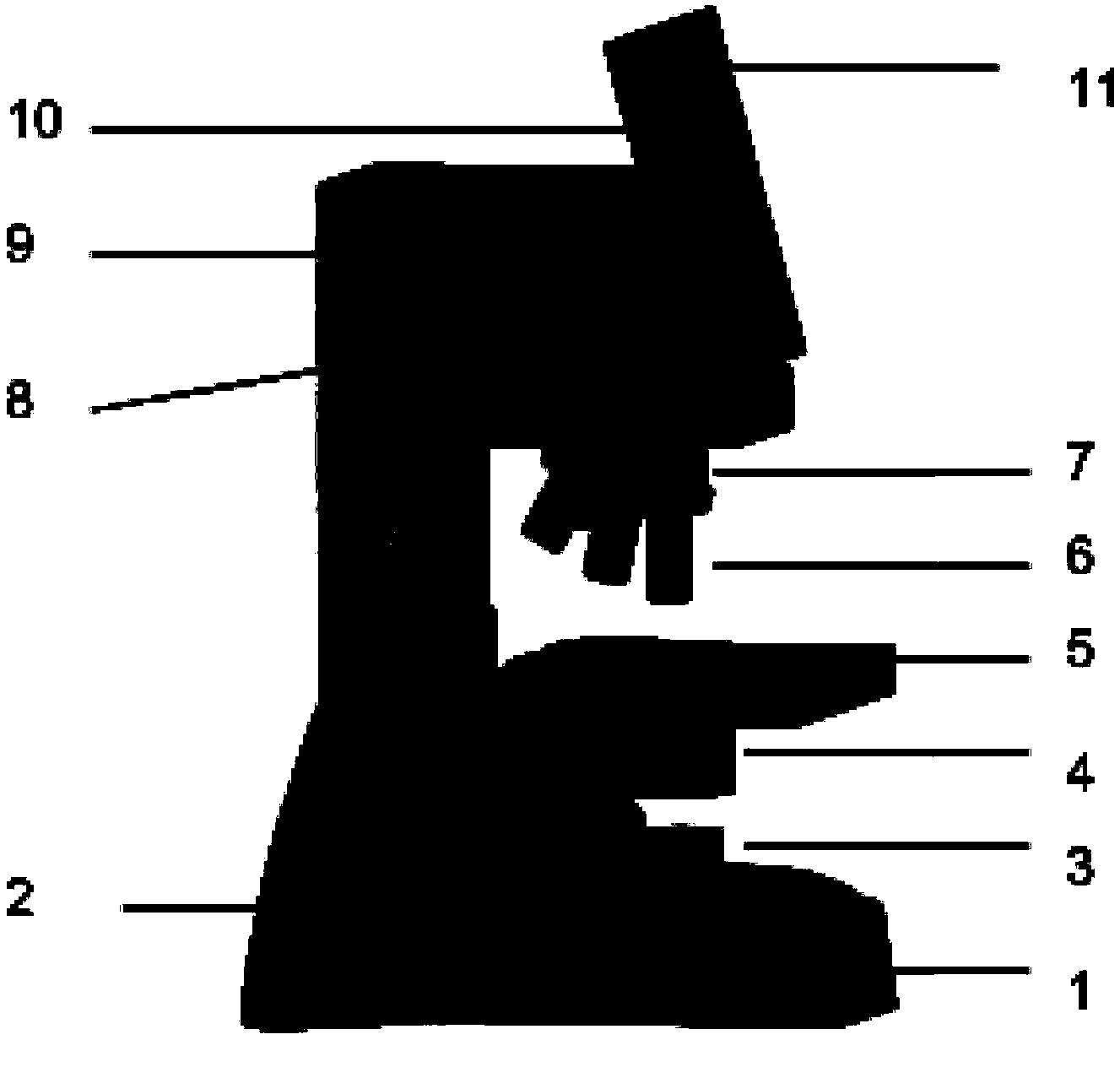
[0037] This embodiment provides a digital microscope for urine sediment detection, its structure is as follows figure 1 shown, including:

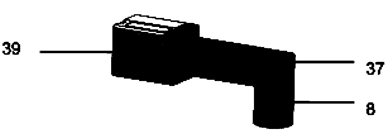
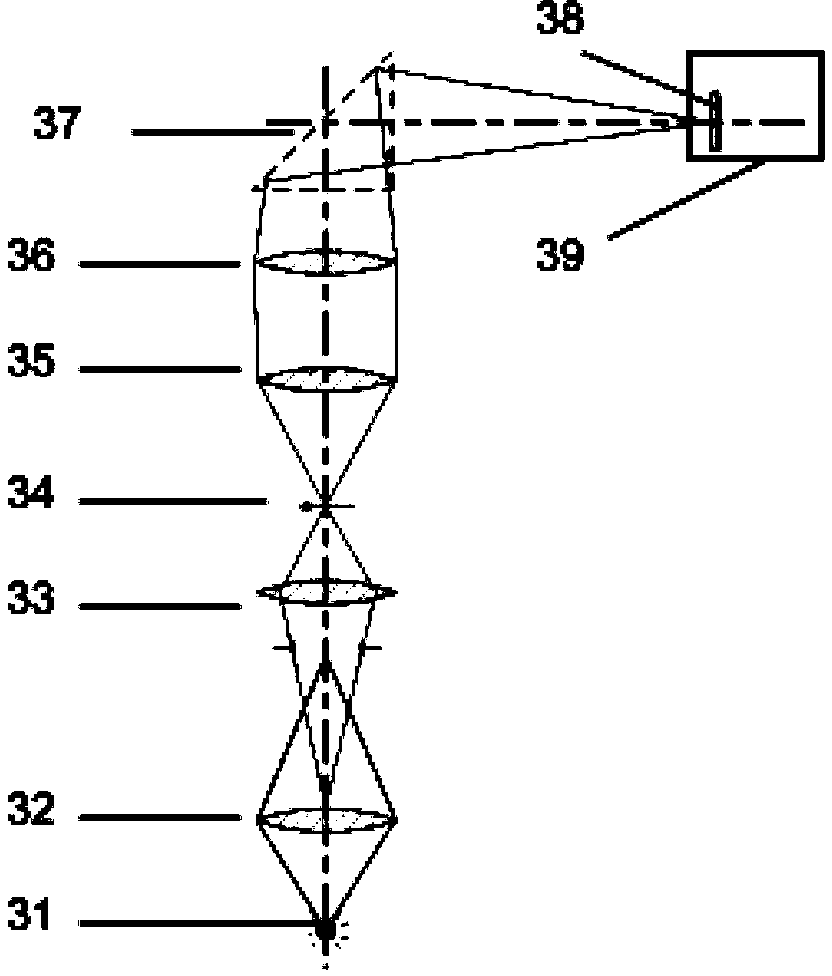
[0038] An upright optical microscope device, which includes from bottom to top: microscope support 1, focusing device 2, lighting device 3, condenser lens 4, sample stage 5, objective lens 6, mirror changer seat 7, lens barrel lens (not shown in the figure) shown), in which the microscope stand 1 is used to support other components of the digital microscope, the illuminating device 3 is located under the sample stage 5, and the condenser lens 4 is located between the illuminating device 3 and the sample stage 5, and is used to convert the light emitted by the illuminating device 3 into Converging to the sample stage 5 to illuminate the urine sediment sample carried therein, the mirror changing rotary seat 7 is located above the sample stage 5, and a plurality of objective lenses 6 with different magnifications are installed on it, and the ...


PUM


Abstract

The invention provides a digital microscope for urinary sediment detection and an image identification method using the digital microscope. The digital microscope comprises an optical microscopic device used for imaging a to-be-detected sample into an amplified image; an imaging device, used for collecting images imaged by the optical microscopic device; a displayer, used for displaying the images collected by the imaging device; and a control device, used to control the imaging device and the displayer and receive image data collected by the imaging device and send the image data to the displayer; wherein the imaging device is provided with an imaging chip, the imaging chip size is 1 / 4 inch-1 inch, and the frame rate is greater than 25 frames per second. The digital microscope is good in imaging effect, accurate in component identification, and low in cost, and can satisfy the wide remote area medical need.

Description

technical field [0001] The invention relates to a digital microscope for urine sediment analysis, in particular to a digital microscope integrated with a display. Background technique [0002] Urine is one of the important excretions in the body, and it is a window to reflect the pathological changes of kidney organs. As an indispensable and important part of complete urinalysis, urine sediment examination is of great significance for the diagnosis, localization, identification, prognosis judgment, drug treatment monitoring and health screening of diseases such as the urinary system. Urinary sediment refers to the shaped components in urine, which is the sediment formed after centrifugation of the original urine. It is a combination of quality and quantity of the formed components of urine, including cells, tubes, crystals, bacteria, sperm, etc. [0003] Traditional manual microscope examination of urine sediment is not only labor-intensive and easy to pollute the environme...

Claims


Application Information

Patent Timeline
IPC IPC(8): G01N15/14G02B21/36
CPCG02B21/24G01N33/493G02B21/0008
Inventor 林科特雷弗·阿利森沈宏邹杰刘宁
Owner SIEMENS HEALTHCARE DIAGNOSTICS INC
Why Eureka
Try Eureka