A thoracoscopic lung fixation device

A fixation device and thoracoscopic technology, applied in the field of medical equipment, can solve problems such as inability to fix the lungs, achieve the effects of avoiding lung movement, adjustable clamping force, and reducing the range of motion

Pending Publication Date: 2019-01-18
SHANGHAI FIRST PEOPLES HOSPITAL
View PDF0 Cites 3 Cited by
  • Summary
  • Abstract
  • Description
  • Claims
  • Application Information

AI Technical Summary

Problems solved by technology

The lung traction forceps can only clamp the lungs during the operation, but cannot fix the lungs under thoracoscopic surgery, and cannot avoid the movement of the lungs caused by the beating of the heart and breathing
In the prior art, there is no report about the thoracoscopic lung fixation device of the present invention

Method used


Image

  • A thoracoscopic lung fixation device
  • A thoracoscopic lung fixation device
  • A thoracoscopic lung fixation device

Examples


Embodiment 1

[0035] Embodiment 1 Thoracoscopic lung fixation device

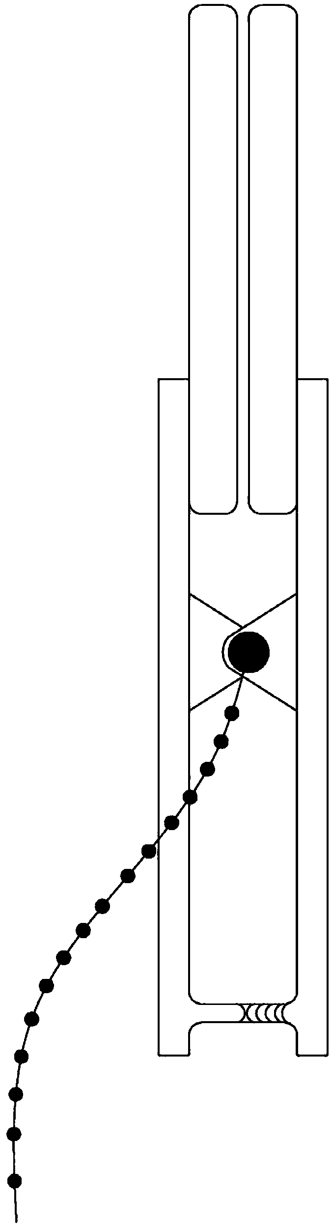
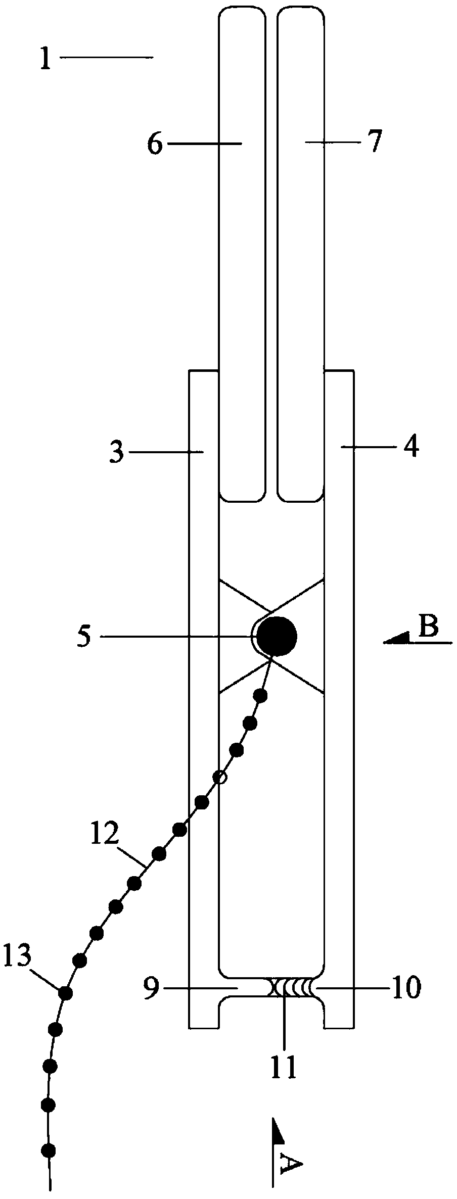
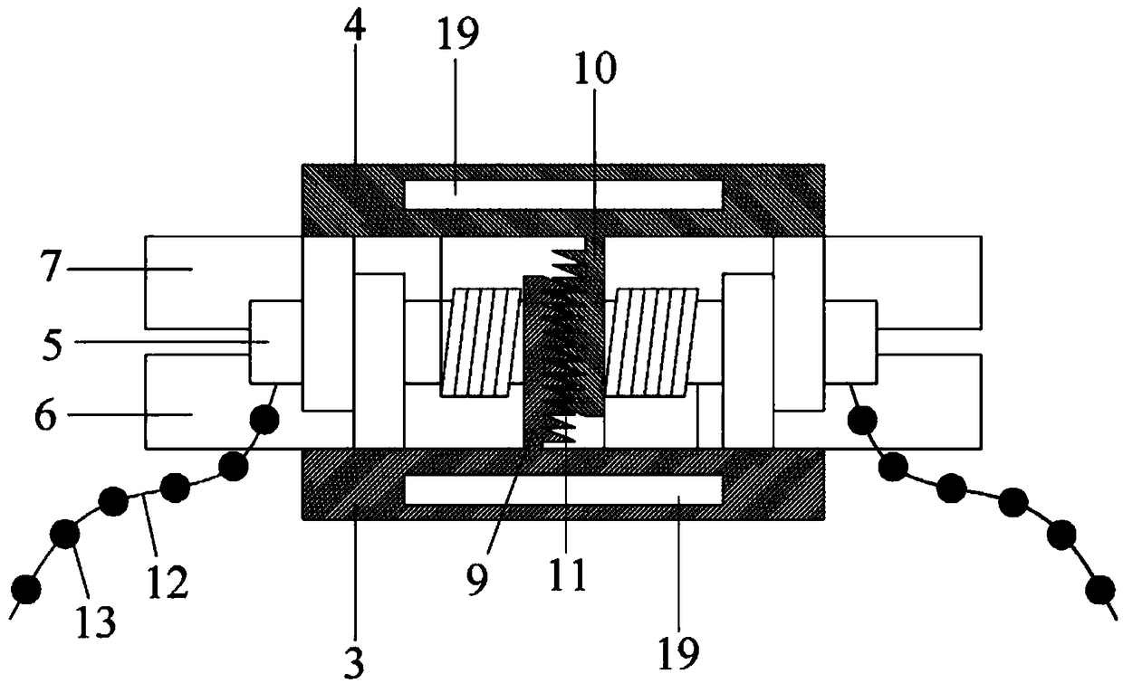
[0036] See Figure 1-Figure 6 . attached figure 1 Schematic diagram of the structure of the lung clamping tool, attached figure 2 Labeled diagram for each part of the lung gripping tool, with image 3 for figure 2 The enlarged schematic diagram of A in the middle, attached Figure 4 for figure 2 The enlarged schematic diagram in the B direction, attached Figure 5 Schematic diagram of the structure of the chest wall fixation device, attached Figure 6 for Figure 5 The enlarged schematic diagram of A in the middle, attached Figure 7 Schematic diagram of the use state for the lung gripping tool.

[0037] The lung fixation device under thoracoscopy of the present invention is composed of two parts, a lung clamping tool 1 and a chest wall fixation device 2. The lung clamping tool 1 is sent into the thoracic cavity under a thoracoscope for clamping the lungs; the chest wall fixation device 2. Penetrate and fix...


PUM


Abstract

The invention relates to the technical field of medical instruments, in particular to a thoracoscopic lung fixation device, comprising a lung clamping tool and a chest wall fixing device. The lung clamping tool comprises left and right clips, and two clips are articulated through a spring column. The head end of the clip is provided with a clip head, the tail end of the clip is provided with a rack, and the left rack and the right rack are provided with fixed clamping teeth capable of engaging with each other. The tail end of the clip is provided with a slot. Suspension wires are respectivelyarranged at the left and right ends of the spring columns, and the suspension wires are spaced at intervals of 1-3 cm position with beads. The chest wall fixing device comprises an elastic pipe, an inner clasp ring and an outer clasp ring respectively fixed at two ends, and a plurality of clasp grooves are arranged on the surface of the outer clasp ring. The device can reduce lung movement causedby beating and breathing of the heart, greatly reduce the range of lung movement during thoracoscopic operation, avoid lung movement from interfering with the operation of a doctor, and provide further guarantee for fine operation and operation safety of the operation.

Description

technical field [0001] The invention relates to the technical field of medical instruments, in particular to a lung fixation device under a thoracoscope. Background technique [0002] Thoracoscopic surgery (video-assisted thoracoscopic surgery) uses modern camera technology and high-tech surgical equipment to complete complex intrathoracic surgery under the chest wall cannula or tiny incision. It has changed the treatment of thoracic surgery diseases. The concept, known as one of the major breakthroughs in the field of thoracic surgery in the last century, is a representative operation of minimally invasive thoracic surgery and the direction of future thoracic surgery development. Complete thoracoscopic surgery only needs to make 1 to 3 small holes of 1.5 cm in the chest wall. The tiny medical camera projects the situation in the chest cavity to the large display screen, which is equivalent to putting the doctor's eyes into the patient's chest cavity for surgery. The surgi...

Claims


Application Information

Patent Timeline
Patent Type & Authority Applications(China)
IPC IPC(8): A61B17/02A61B17/29
CPCA61B17/0218A61B17/29A61B2017/2926
Inventor 邓国英胡海洋李春霖王琦张永兴
Owner SHANGHAI FIRST PEOPLES HOSPITAL
Why Eureka
Try Eureka