A method and system for image segmentation of intracranial arteries

A technology of arterial blood vessels and images, which is applied in the field of medical imaging, can solve the problems of unclear edges and time-consuming filtering of intracranial arterial blood vessel images, and achieve the effects of saving time-consuming filtering, clear edges, and accurate segmentation

Active Publication Date: 2021-03-02
UNION STRONG (BEIJING) TECH CO LTD
View PDF4 Cites 0 Cited by
  • Summary
  • Abstract
  • Description
  • Claims
  • Application Information

AI Technical Summary

Problems solved by technology

[0004] The embodiment of the present application provides a method and system for segmenting intracranial arteries and blood vessels, which are used to solve the problem of unclear edges of intracranial arteries and blood vessels in the segmentation of three-dimensional CTA images, as well as the time-consuming problem of filtering, thereby realizing accurate three-dimensional CTA images segmentation

Method used


Image

  • A method and system for image segmentation of intracranial arteries
  • A method and system for image segmentation of intracranial arteries
  • A method and system for image segmentation of intracranial arteries

Examples


Embodiment Construction

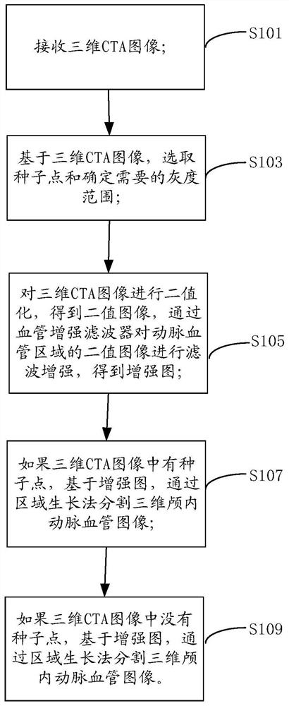
[0032] Embodiments of the present application provide a method and system for segmenting intracranial blood vessel images, which are used to solve the problem of unreliable segmentation techniques for intracranial artery and blood vessel images in three-dimensional CTA images.

[0033] see figure 1 , a method for image segmentation of intracranial arteries, comprising:

[0034] S101: receiving a three-dimensional CTA image;

[0035] S103: Based on the three-dimensional CTA image, select the seed point and determine the required gray scale range;

[0036] S105: Perform binarization on the three-dimensional CTA image to obtain a binary image, and enhance the binary image of the intracranial artery and blood vessel image area through a blood vessel enhancement filter to obtain an enhanced image;

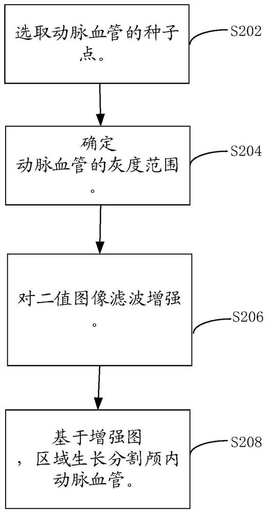
[0037] S107: If there is a seed point in the three-dimensional CTA image, based on the three-dimensional CTA image, segment the three-dimensional intracranial artery image by a region...


PUM


Abstract

The embodiment of the present application discloses a method and system for segmenting an intracranial artery image. The solution includes: receiving a 3D CTA image; selecting seed points and determining the required gray scale range based on the 3D CTA image; binarizing the 3D CTA image to obtain a binary image; The binary image of the 3D CTA image is enhanced to obtain an enhanced map; if there are seed points in the 3D CTA image, based on the enhanced map, the 3D intracranial artery image is segmented by the region growing method; if there is no seed point in the 3D CTA image, based on the enhanced map, by Segmentation of three-dimensional intracranial arterial images by interval binary segmentation. This scheme makes the edge of the segmented three-dimensional intracranial arterial image clear, saves time-consuming filtering, and thus realizes accurate segmentation of the intracranial arterial image of the three-dimensional CTA image.

Description

technical field [0001] The present application relates to the field of medical imaging, in particular to a method and system for segmenting images of intracranial arteries. Background technique [0002] With the continuous progress and development of modern medical imaging equipment, the development of medical imaging technology and computer graphics has brought great changes to medicine. Computed tomography (CT), nuclear magnetic resonance (MRI), ultrasound (US), positron Emission computed tomography (PET), CTA and other medical imaging technologies have been widely used in clinical diagnosis. CTA is an angiographic examination using computer-assisted tomography (CT) technology, which can explore all arteries and veins in the body, including blood vessels in the heart, brain, lungs, kidneys, and limbs. Since Rontgen discovered X-rays, people can use X-ray perspective equipment to detect information inside objects. Its technology mainly makes blood less permeable to X-rays...

Claims


Application Information

Patent Timeline
Patent Type & Authority Patents(China)
IPC IPC(8): G06T7/11
CPCG06T2207/10081G06T2207/20156G06T2207/20192G06T2207/30101G06T7/11
Inventor 杨光明冯雪王文智秦岚
Owner UNION STRONG (BEIJING) TECH CO LTD
Why Eureka
Try Eureka