A method for segmenting human organs in medical images based on a neural network

A medical imaging and neural network technology, applied in image analysis, instruments, image enhancement, etc., can solve the problems of inability to achieve accurate positioning of the initial layer and the end layer of blood vessels, damage to the Z axis, and small differences, etc., to solve multiple problems. Effects on the organ segmentation problem

Pending Publication Date: 2019-03-12
BEIJING LINKING MEDICAL TECH CO LTD
View PDF0 Cites 5 Cited by
  • Summary
  • Abstract
  • Description
  • Claims
  • Application Information

AI Technical Summary

Problems solved by technology

(1). If data enhancement is not performed, it is easy to cause data imbalance between organs; (2). The HU values ​​​​of each organ corresponding to different regions are close to each other, and there is little difference from each other. It is easy to confuse and segment the three; ( 3). If data enhancement is performed, the Z-axis will be destroyed (the Z-axis refers to the upper and lower layers of the medical image. For example, if a person is 170cm tall, if the corresponding layer thickness between CT slices is 5mm when taking a CT scan, the total There are 1700 / 5=340 pieces of CT, where 340 corresponds to the information of the Z axis), the idea of ​​using a 3D U-shaped neural network to perform fine segmentation after rough positioning will fail, and the existing rough positioning of the human body The multi-classification network cannot realize the precise positioning of the initial layer and the end layer of the blood vessel

Method used


Image

  • A method for segmenting human organs in medical images based on a neural network
  • A method for segmenting human organs in medical images based on a neural network
  • A method for segmenting human organs in medical images based on a neural network

Examples


Embodiment 1

[0035] A method for segmenting human organs in medical images based on a neural network, suitable for execution in a computing device, comprising the following steps:

[0036] (1) Coarsely locate the medical imaging layer where the target organ to be delineated is located; the target organ to be delineated includes several organs; preferably further comprising the following steps:

[0037] (1a) Divide the organs in the medical image into several categories according to their high and low positions;

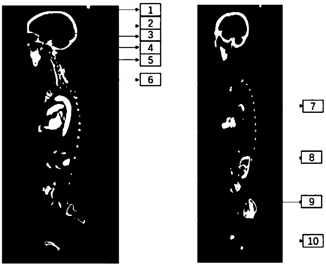
[0038] in such as figure 1 In the exemplary embodiment shown in Table 1, the organs in the human body medical image are classified into ten categories from top to bottom or from bottom to top, which are from the first picture to the top of the head, from the top of the head to the top layer of the eye, From the upper roof of the eye to the lower roof of the eye, from the lower roof of the eye to the lower roof of the cerebellum, from the lower roof of the cerebellum to the last l...

Embodiment 2

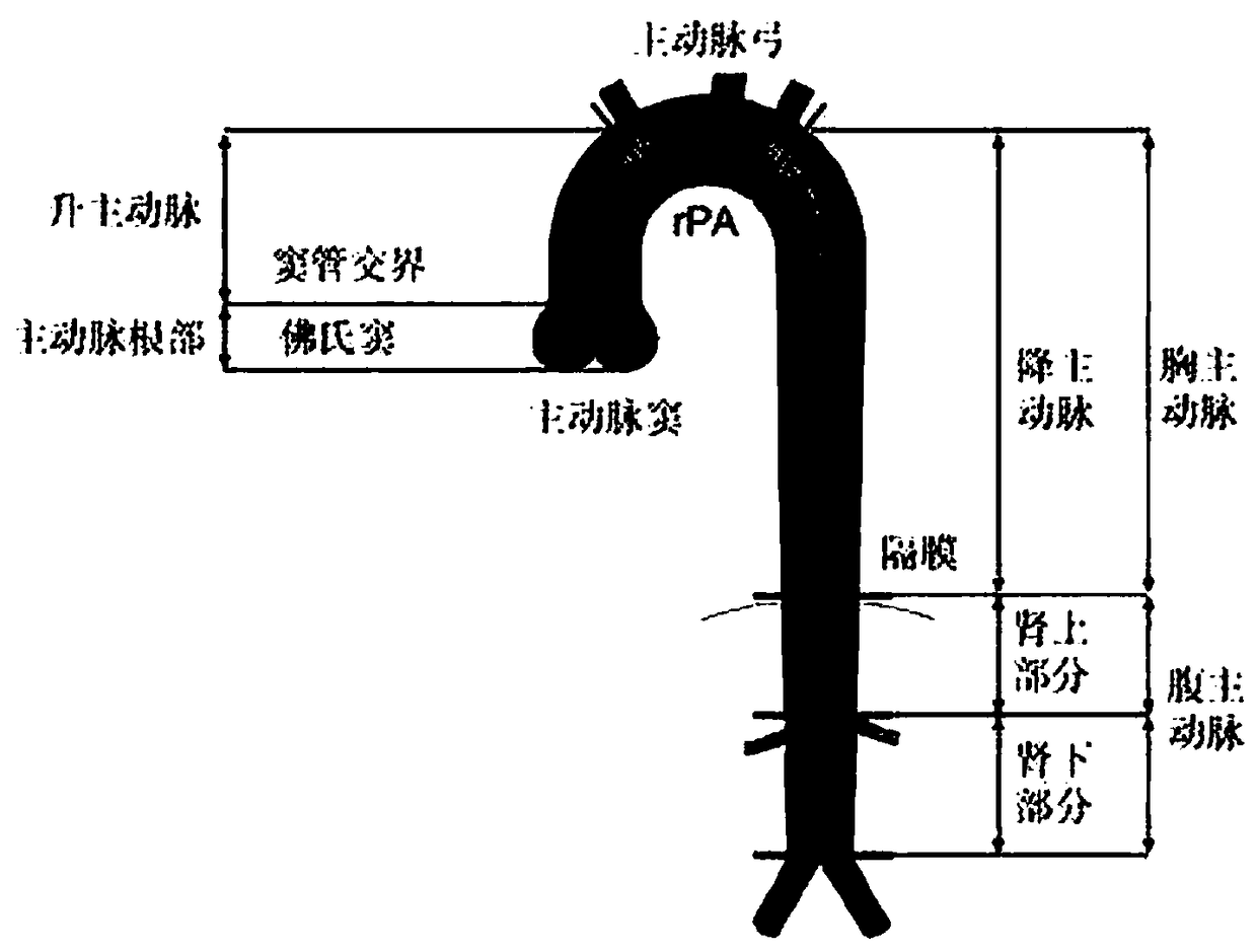
[0049] In an exemplary embodiment, for example, in organ segmentation of medical images, especially in the segmentation of slender organs like blood vessels, the regions corresponding to different names often have different lengths, and it is easy to cause differences in data between categories during training. Balance, such as the ascending aorta, aortic arch, and descending aorta, these three vessels (organs) are on the same vessel (see figure 1 ), have different names because of the location of the area. These three blood vessels are respectively located in the [7,8,9] layer of the multi-classification localization network of the human body (as shown in Table 1). The descending aorta is the longest, and the ascending aorta is the longest. The arteries are next, and the aortic arch is the shortest. Specifically, taking a CT of a patient as an example, when the Z-axis spacing (spacing) is 3 mm, that is, when the CT slice thickness is 3 mm, there are 23 slices of the ascending...

Embodiment 3

[0058] The invention also provides a computing device, comprising:

[0059] one or more processors;

[0060] storage; and

[0061] One or more programs, wherein the above one or more programs are stored in the above memory and configured to be executed by one or more processors, the above one or more programs include a method for performing neural network-based analysis of the human body in medical images Instructions for a method for segmenting an organ, the method comprising the steps of:

[0062] (1) roughly locating the medical image layer where the target organ to be delineated is located; the target organ to be delineated includes several organs;

[0063] (2) Determine the start layer and end layer of all target organs to be delineated as a whole;

[0064] (3) Input the middle layer of the start layer and the end layer into the 2D multi-classification U-shaped network, perform fine delineation and segmentation, and determine the corresponding start layer and end layer...


PUM


Abstract

The invention belongs to the technical field of depth learning and radiotherapy, and relates to a method for segmenting human organs in medical images based on a neural network. The method comprises the following steps: roughly positioning a medical image layer where a target organ to be delineated is located, wherein the target organ to be delineated comprises a plurality of organs; A 3D binary U-shaped network is used to determine the initial and final layers of the target organ to be delineated as a whole. The middle layer of the beginning layer and the end layer is input into a 2D multi-class U-shaped network for fine delineation and segmentation, and the corresponding beginning layer and the end layer of each organ to be delineated are determined. The present invention uses a 3D network to determine a starting layer and an ending layer when multiple organs are integrated, A 2D network is use to solve that data imbalance problem between the organs between the start lay and the endlayer, and by combining the advantages of the existing 3D network and the 2D network, the problem of segmentation of multiple organ with similar HU values in the prior art is solved.

Description

technical field [0001] The invention belongs to the technical field of deep learning and radiotherapy, and relates to a method, device and storage medium for segmenting human organs in medical images by combining a 2D multi-classification U-shaped neural network and a 3D binary classification U-shaped neural network. Background technique [0002] As we all know, the human body contains a variety of slender organs. For example, the segmentation of blood vessels usually faces some difficulties: in the same set of CT images of the same patient, the HU values ​​​​of the blood vessels are generally similar, but the blood vessels in different regions have different names. It brings trouble to the image segmentation of blood vessels. (1). If data enhancement is not performed, it is easy to cause data imbalance between organs; (2). The HU values ​​​​of each organ corresponding to different regions are close to each other, and there is little difference from each other. It is easy to...

Claims


Application Information

Patent Timeline
Patent Type & Authority Applications(China)
IPC IPC(8): G06T7/11G06K9/62
CPCG06T7/11G06T2207/30004G06T2207/30101G06T2207/20081G06T2207/20084G06T2207/10081G06F18/2431G06F18/2433
Inventor 胡志强武会杰崔德琪章桦
Owner BEIJING LINKING MEDICAL TECH CO LTD
Why Eureka
Try Eureka