J-subgroup avian leukosis virus (ALV) antibody quick detection test strip and preparation method and application thereof

An avian leukemia virus and detection test strip technology, which is applied in biological tests, measuring devices, material inspection products, etc., can solve the problems of false positives, false negatives, low specificity, etc., and achieves fast and simple preparation and sample detection, convenient operation, The effect of high antigen purity

Inactive Publication Date: 2019-06-07
YANGZHOU UNIV
View PDF5 Cites 0 Cited by
  • Summary
  • Abstract
  • Description
  • Claims
  • Application Information

AI Technical Summary

Problems solved by technology

At present, there are not many methods for detecting ALV-J subgroup-specific antibodies, and these kits are unstable and have low specificity, and false positives and false negatives often occur, such as: commercial ALV-J antibody ELISA detection on the market Reagent test kit

Method used


Image

  • J-subgroup avian leukosis virus (ALV) antibody quick detection test strip and preparation method and application thereof
  • J-subgroup avian leukosis virus (ALV) antibody quick detection test strip and preparation method and application thereof
  • J-subgroup avian leukosis virus (ALV) antibody quick detection test strip and preparation method and application thereof

Examples


Embodiment 1

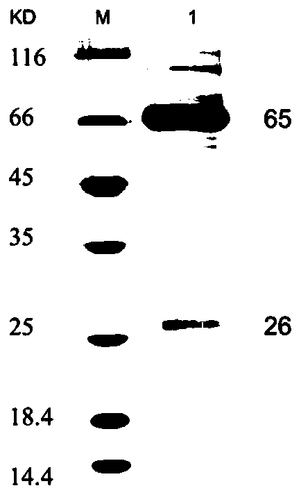
[0027] Chicken IgG with high purity was obtained by saturated ammonium sulfate precipitation method and sephadex gel purification method. The specific process is:

[0028] (1) Take 1mL of chicken serum, centrifuge at 10000RPM for 5min, and remove sediment and floating matter.

[0029] (2) Transfer the supernatant obtained by centrifugation to a 10mL centrifuge tube, then add 3mL PBS, and mix gently on a shaker in an ice bath.

[0030] (3) Add 4 mL of saturated ammonium sulfate (pH=7.4) dropwise to make the final concentration of ammonium sulfate 50%, and place it at 4°C for 30 minutes.

[0031] (4) Divide the mixture into centrifuge tubes, centrifuge at 11,000 RPM at 4°C for 20 min.

[0032] (5) Discard the supernatant after centrifugation, resuspend the precipitate to 5 mL with PBS (each finger tube is washed 3 times with 200 μL PBS, transfer the washing solution to a centrifuge tube, add PBS to 5 mL), slowly add saturated sulfuric acid dropwise Ammonium 2.7mL, so that the...

Embodiment 2

[0038] Embodiment 2 Establishment of Monoclonal Antibody Hybridoma Cell Line

[0039] Five 6-8 week old BALB / C female mice were immunized with purified chicken IgG protein. For the first immunization, 100 μg was mixed with an equal volume of complete Freund’s adjuvant, and injected intraperitoneally; for the second immunization, 200 μg was mixed with an equal volume of incomplete Freund’s adjuvant, and injected intraperitoneally; for the third immunization, each mouse was 200 μg, without adjuvant , intraperitoneal injection. The interval between each immunization was 15 days, and the immunization was boosted once 3 days before the fusion. Take mouse splenocytes and myeloma cells (SP2 / 0) in the logarithmic growth phase for cell fusion at a ratio of 1:7, and use 50% PEG4000 for fusion. Cultured in HAT medium containing 15% fetal bovine serum, adding appropriate amount of ICR mouse peritoneal macrophages as feeder cells. After 5 days of fusion, the medium was completely change...

Embodiment 3

[0045] Colloidal gold antibody preparation

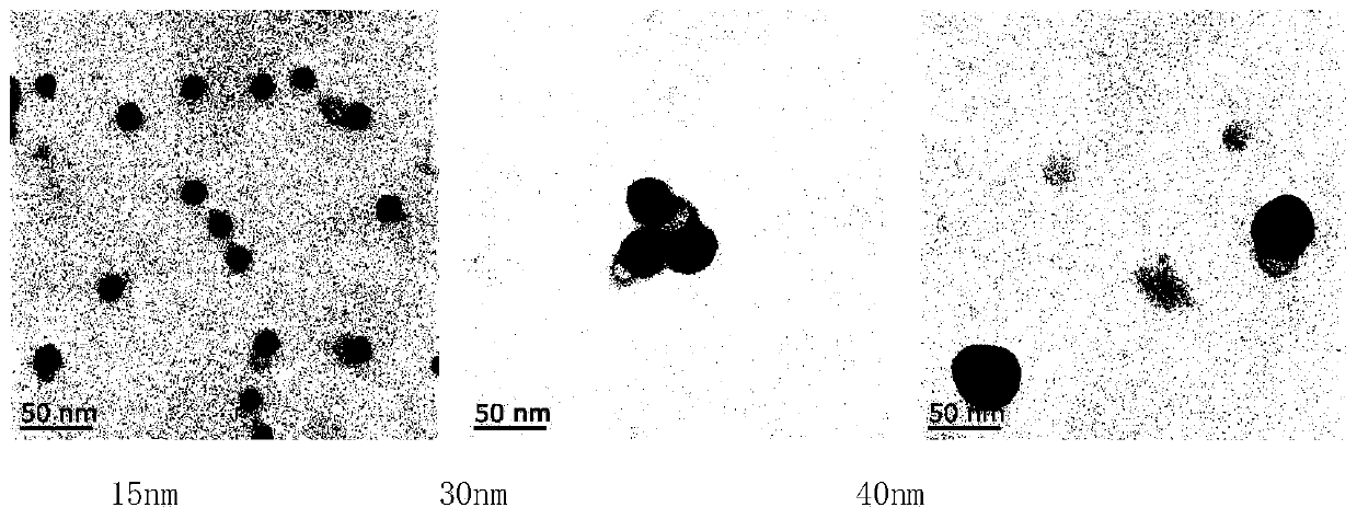
[0046] with 0.1M K 2 CO 3 The pH of the colloidal gold solution of three different particle sizes (40nm, 30nm, 15nm) was adjusted to 8.8, and 0, 1, 2, 3, 4, 5, 6, 7 μg of single Anti-cIgG-5B3 (that is, the monoclonal antibody expressed by the hybridoma cell line cIgG-5B3 that has been biologically preserved), reacted at room temperature for 15 minutes, added 100 μL of 10% NaCl solution, mixed well and stood at room temperature for 20 minutes. The minimum stable amount, 10% more than the minimum stable amount, is the optimal amount of protein required for labeling.

[0047] Take three kinds of colloidal gold solutions with different particle sizes (40nm, 30nm, 15nm) and observe them under light. As the particle size increases, the color of the colloidal gold solution gradually becomes darker. Colloidal gold solution bottom all has no conglomerate, and light transmittance is good; Observation under transmission electron microscope ...


PUM


Abstract

The invention relates to a J-subgroup avian leukosis virus (ALV) antibody quick detection test strip and a preparation method and application thereof. The ALV-J envelope protein gp85 specific epitopeserves as an antigen to detect an ALV-J specific antibody in serum; the specific polypeptide epitope serves as an antigen to be spray-painted on an NC film, and a detection line of the test strip is made; and a monoclonal mouse anti-chicken IgG antibody is selected as a gold-labelled antibody, and a prepared colloidal gold-antibody coupled composite is sprayed on a gold-labelled pad of the test strip. The antigen in the serum of ALV-J artificially infected chickens can be detected through the test strip; and the test strip can be used for evaluating whether ALV-J infection exists in a chickengroup or not, and is easy and convenient to prepare, low in cost, wide in application and high in economic benefit.

Description

technical field [0001] The invention relates to a test strip for rapid detection of J subgroup avian leukosis virus antibody, a preparation method and application thereof. Background technique [0002] Subgroup J avian leukemia is a contagious tumor disease of poultry caused by subgroup J avian leukemia virus (ALV-J). ALV-J has brought huge economic losses to the poultry industry due to the influence of factors such as induced tumors, discarded chicken carcasses, and decreased egg production performance on the performance of chicken flocks. At present, there is no effective treatment and vaccine for subgroup J avian leukemia, and it can only be controlled by specific diagnosis, elimination of positive chickens and population purification. The medium and long-term plan of the Ministry of Agriculture of my country has clearly defined the purification work of avian leukosis in the chicken industry. The evaluation of decontaminated flocks often uses the method of antigen antib...

Claims


Application Information

Patent Timeline
Patent Type & Authority Applications(China)
IPC IPC(8): G01N33/569G01N33/574G01N33/68G01N33/577G01N33/558
Inventor 秦爱建沈习赵巍钱琨邵红霞叶建强
Owner YANGZHOU UNIV
Why Eureka
Try Eureka