Respiratory signal extraction method and device for contrast image sequence

A technology for contrast images and respiratory signals, which is applied in the field of image processing and can solve problems such as unfavorable real-time operations

Active Publication Date: 2021-05-14
BEIJING INSTITUTE OF TECHNOLOGYGY
View PDF10 Cites 0 Cited by
  • Summary
  • Abstract
  • Description
  • Claims
  • Application Information

AI Technical Summary

Problems solved by technology

They all have certain limitations: In the first category of methods, the region of interest is the diaphragm structure
The limitation of this method is that the specific dimensionality reduction modeling process needs to input a sequence of images first, which is not conducive to real-time operation during surgery

Method used


Image

  • Respiratory signal extraction method and device for contrast image sequence
  • Respiratory signal extraction method and device for contrast image sequence
  • Respiratory signal extraction method and device for contrast image sequence

Examples


Embodiment Construction

[0028] In order to make the purpose, technical solutions and advantages of the embodiments of the present invention clearer, the technical solutions in the embodiments of the present invention will be clearly and completely described below in conjunction with the drawings in the embodiments of the present invention. Obviously, the described embodiments It is a part of embodiments of the present invention, but not all embodiments. Based on the embodiments of the present invention, all other embodiments obtained by persons of ordinary skill in the art without creative efforts fall within the protection scope of the present invention.

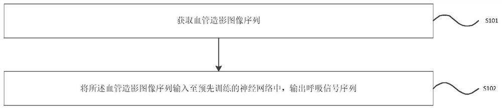
[0029] figure 1 It is a schematic flow chart of the respiratory signal extraction method of the contrast image sequence according to the embodiment of the present invention, as shown in figure 1 As shown, the method includes:

[0030] S101. Acquire a contrast image sequence. The contrast image sequence in the embodiment of the present invention...


PUM


Abstract

An embodiment of the present invention provides a method and device for extracting respiratory signals of a contrast image sequence, wherein the method includes: acquiring a contrast image sequence, inputting the contrast image sequence into a pre-trained neural network, and outputting a respiratory signal sequence; the neural network includes a self-encoder The device layer is used to input the contrast image sequence and output the background image of each frame image in the contrast image sequence to form the background image sequence; the optical flow network layer is used to input the background image sequence and output the motion of the background image sequence Information; a recurrent neural network layer, used to input the motion information of the background image sequence and output the respiratory signal sequence. The embodiment of the present invention can automatically and accurately extract the respiratory signal sequence.

Description

technical field [0001] Embodiments of the present invention relate to the technical field of image processing, and more specifically, to a method and device for extracting respiratory signals of a contrast image sequence. Background technique [0002] Angiography is an interventional detection method in which a contrast agent is injected into blood vessels. Because X-rays cannot penetrate the contrast agent, the image displayed by the contrast agent under X-ray is considered to be a blood vessel. Currently, contrast images are the most common imaging modality in thoracic and abdominal vascular interventional procedures. In thoracic and abdominal related operations, the patient's breathing will cause the displacement of various tissues and organs, so in the image, the position of the contrast agent will also change, which will bring difficulties to the doctor's positioning operation. In order to compensate the displacement of tissues and organs in the image with respiration,...

Claims


Application Information

Patent Timeline
Patent Type & Authority Patents(China)
IPC IPC(8): G06T7/00G06T7/194G06T7/269G06K9/00G06N3/04G06N3/08
CPCG06T7/0012G06T7/194G06T7/269G06N3/08G06T2207/10116G06T2207/30101G06T2207/20084G06T2207/20081G06V2201/03G06N3/045G06F2218/12
Inventor 王涌天杨健艾丹妮范敬凡方慧卉
Owner BEIJING INSTITUTE OF TECHNOLOGYGY
Why Eureka
Try Eureka