

Combined detection method for pepsinogen I and pepsinogen II
A pepsinogen and combined detection technology, applied in the field of in vitro diagnosis, can solve problems such as high inspection costs, high technical requirements for operators, and radiation exposure, and achieve the effect of avoiding human body damage, avoiding the inconvenience of gastroscopy, and good correlation
- Summary
- Abstract
- Description
- Claims
- Application Information
AI Technical Summary
Problems solved by technology
Method used
Image

Examples
Embodiment 1
[0023] The combined detection method of pepsinogen Ⅰ and pepsinogen Ⅱ comprises the following steps:
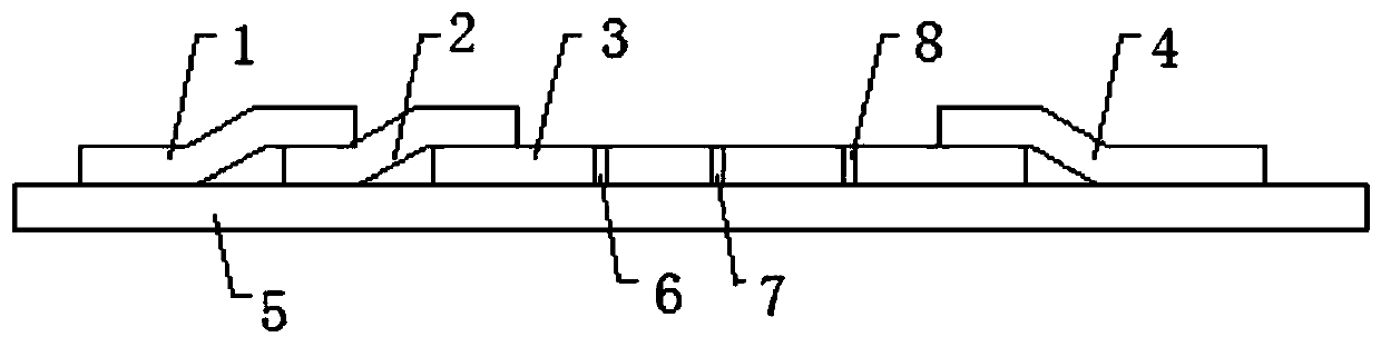
[0024] (1) Immunochromatography test strip assembly: Lap the sample pad, binding pad, reaction membrane and water-absorbent pad sequentially and paste them on the base plate. The thickness of the base plate is 0.2 mm, the length is 7 cm, and the width is 4 mm. A detection line I and a detection line II are also arranged between the line and the bonding pad, and the detection line II is located between the detection line I and the control line,
[0025] (2) Antibody coating: fluorescent latex-labeled pepsinogen Ⅰ monoclonal antibody with a concentration of 0.4 mg / ml, fluorescent latex-labeled pepsinogen Ⅱ monoclonal antibody with a concentration of 0.4 mg / ml and a concentration of 0.4 mg / ml of mouse anti-rabbit IgG coated on the binding pad, the concentration of 0.8mg / ml pepsinogen Ⅰ monoclonal antibody, the concentration of 0.8mg / ml pepsinogen Ⅱ monoclonal antibody and the c...
Embodiment 2
[0029] The combined detection method of pepsinogen Ⅰ and pepsinogen Ⅱ comprises the following steps:
[0030] (1) Immunochromatography test strip assembly: Lap the sample pad, binding pad, reaction membrane and water-absorbent pad sequentially and paste them on the bottom plate. The thickness of the bottom plate is 0.3mm, the length is 6cm, and the width is 4mm. A detection line I and a detection line II are also arranged between the line and the bonding pad, and the detection line II is located between the detection line I and the control line,
[0031] (2) Antibody coating: fluorescent latex-labeled pepsinogen Ⅰ monoclonal antibody with a concentration of 0.4 mg / ml, fluorescent latex-labeled pepsinogen Ⅱ monoclonal antibody with a concentration of 0.4 mg / ml and a concentration of 0.4 mg / ml of mouse anti-rabbit IgG coated on the binding pad, the concentration of 0.8mg / ml pepsinogen Ⅰ monoclonal antibody, the concentration of 0.8mg / ml pepsinogen Ⅱ monoclonal antibody and the ...
Embodiment 3
[0035] The present invention also includes a preparation method of fluorescent latex-labeled pepsinogen I monoclonal antibody:
[0036] Add 2% BSA to a 0.05MPBS solution with a pH value of 7.5 and store at 4°C to prepare a labeled washing solution; add 50mg / ml EDC solution to a 0.05MPBS solution buffer with a pH value of 7.5 to prepare a latex crosslinking agent. Mix 1% BSA, 0.5% skimmed milk, 0.05% NaN3, 0.1% Tween-20 and 0.01MPBS solution with a pH value of 7.5 to prepare a latex preservation solution; After reacting at room temperature for 1 hour, centrifuge for 15 minutes, centrifuge 3 times with 0.05 MPBS to obtain activated fluorescent latex; add 0.4 mg of pepsinogen I monoclonal antibody per ml of activated fluorescent latex, mix and stir at room temperature React for 2-4 hours, centrifuge for 30 minutes, discard the supernatant, wash the precipitate twice with labeled washing solution, discard the supernatant for the last time, dissolve the precipitate with latex prese...
PUM
| Property | Measurement | Unit |
|---|---|---|
| Thickness | aaaaa | aaaaa |
| Length | aaaaa | aaaaa |
| Width | aaaaa | aaaaa |
Abstract
Description
Claims
Application Information