Rheumatoid arthritis scoring system based on multi-mode photoacoustic/ultrasonic imaging and application of rheumatoid arthritis scoring system

A rheumatoid, ultrasonic imaging technology, applied in the field of medical diagnosis, can solve the problems of limited clinical use, large size of PAT instruments, complicated settings, etc., and achieve the effect of simple and easy implementation process and calculation process

Inactive Publication Date: 2019-12-27
PEKING UNION MEDICAL COLLEGE HOSPITAL CHINESE ACAD OF MEDICAL SCI
View PDF0 Cites 7 Cited by
  • Summary
  • Abstract
  • Description
  • Claims
  • Application Information

AI Technical Summary

Problems solved by technology

However, the PAT instrument has a large volume, complicated settings,

Method used


Image

  • Rheumatoid arthritis scoring system based on multi-mode photoacoustic/ultrasonic imaging and application of rheumatoid arthritis scoring system
  • Rheumatoid arthritis scoring system based on multi-mode photoacoustic/ultrasonic imaging and application of rheumatoid arthritis scoring system
  • Rheumatoid arthritis scoring system based on multi-mode photoacoustic/ultrasonic imaging and application of rheumatoid arthritis scoring system

Examples


Embodiment 1

[0049] Example 1 Construction of Multimodal Photoacoustic / Ultrasonic Imaging System

[0050] 1. Data analysis and image acquisition

[0051] (1) Inspection procedure

[0052] Select MCP 2, MTP 3, PIP 2, PIP 3, MTP 2, MTP 5 and the wrist on the clinically dominant side for multimodal imaging. Prepare a white flat surface for the patient and place it on the table next to the imaging system. Place the probes on the dorsal, palm or lateral (MCP 2 and MTP 5) sides of the fingers and wrist with the gel sandwiched between them. First, traditional ultrasound scans of joints, including grayscale ultrasound, CDUS, and PDUS, are performed by an experienced ultrasound operator. Afterwards, PA / US imaging was performed on each joint by the same operator. By clipping the PA button on the operating screen, the laser is turned on and live PA and US imaging are played simultaneously. The depth for visualizing the MCP and PIP joints was set at 2 cm, and the depth at the wrist was 2.5-3 cm. ...

Embodiment 2

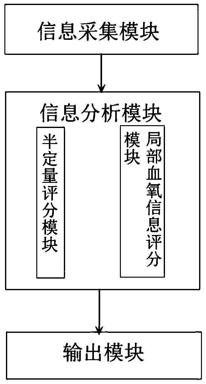
[0071] Example 2. The scoring system for rheumatoid arthritis based on multimodal photoacoustic / ultrasonic imaging such as image 3 as shown,

[0072] A scoring system for rheumatoid arthritis based on multimodal photoacoustic ultrasound imaging, including an information collection module, an information analysis module, and an output module,

[0073] The information collection module uses photoacoustic / ultrasound dual-modal imaging to collect image information of joints in vitro, so as to obtain image information of local inflammatory areas of rheumatoid joints;

[0074] The information analysis module classifies and calculates the collected image information to obtain the characteristic parameters of the image;

[0075] The judgment output module judges the disease activity of the rheumatoid arthritis patient and outputs the data in combination with the characteristic parameters of the image.

[0076] The information analysis module includes a semi-quantitative scoring mo...

Embodiment 3

[0079] Embodiment 3, scoring application example

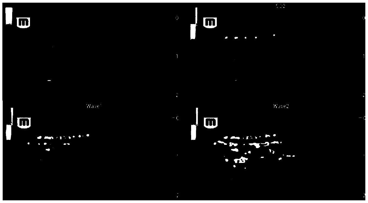
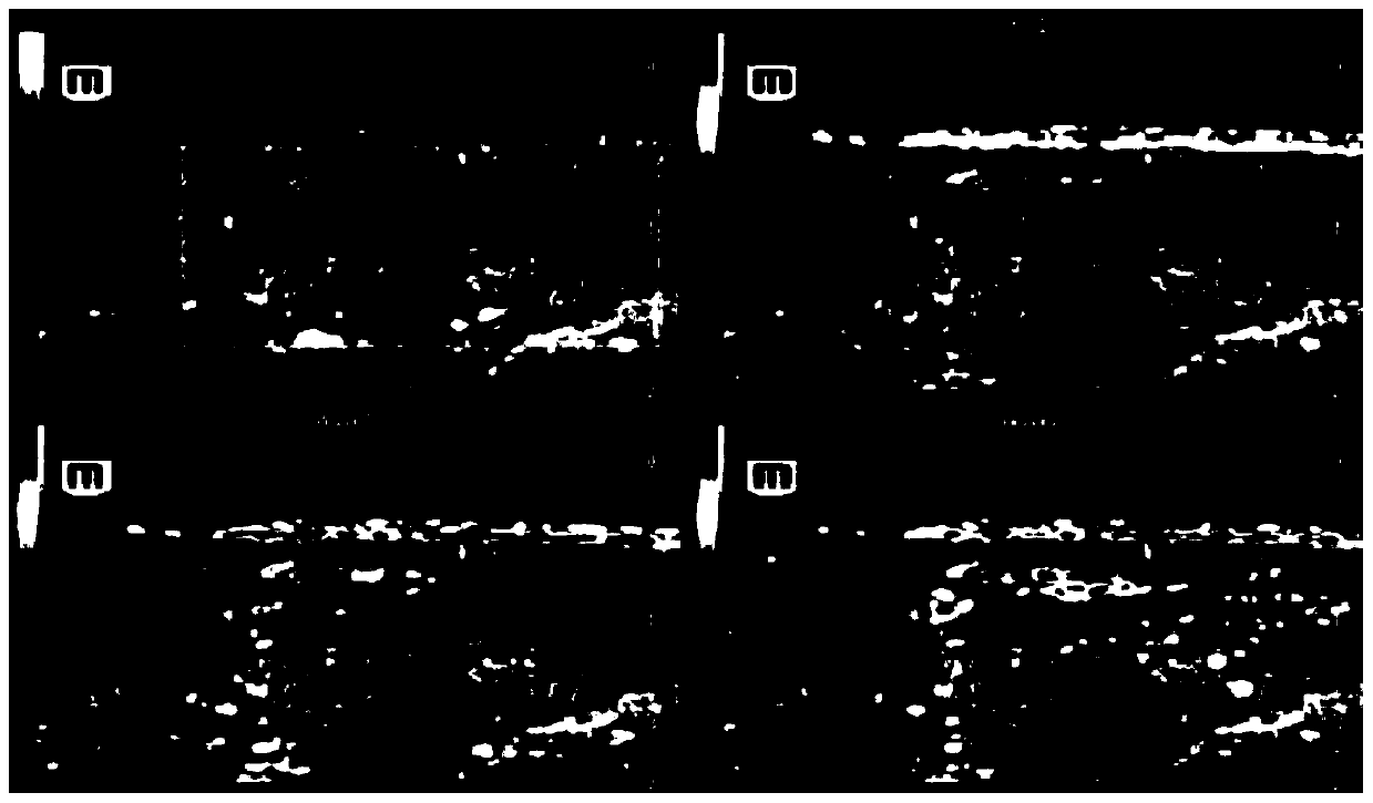
[0080] In April 2019, a 45-year-old female patient clinically diagnosed with RA was selected for multimodal imaging examination. A total of 7 joints on the clinically obvious side of the patient were examined. There was no obvious blood flow signal in the MTP2, MTP3, MCP2, MCP3, PIP2, and PIP3 joint synovium, tendon sheath, and PD / PA in the surrounding area. The imaging findings of the wrist joint were as follows: Figure 4 shown. The wrist joint synovium was obviously thickened, and color Doppler (PD) showed blood flow signals in the thickened area. Both sonographers scored 2 points. In the images of two wavelengths of photoacoustic (PA), more signals were selected The 830nm image of the patient was evaluated, and both doctors scored 3 points. The patient's multimodal imaging color Doppler (PD) total score was 2 points, and the photoacoustic (PA) total score was 3 points, and the blood flow signal Divided into the red grou...


PUM


Abstract

The invention relates to a rheumatoid arthritis scoring system based on multi-mode photoacoustic/ultrasonic imaging and an application of the rheumatoid arthritis scoring system. The application of multi-mode photoacoustic/ultrasonic imaging in the rheumatoid arthritis scoring system is characterized by comprising the following steps: (1) carrying out image information acquisition on joints through photoacoustic/ultrasonic bimodal imaging in an in-vitro mode; (2) analyzing the acquired image information and clinical data to carry out multi-mode photoacoustic/ultrasonic scoring; and (3) judgingdisease activity of rheumatoid arthritis patients according to the multi-mode photoacoustic/ultrasonic scoring combined with local blood oxygen information. The multi-mode photoacoustic/ultrasonic imaging system disclosed by the invention is used for evaluating rheumatoid arthritis for the first time, and has the advantages that a handheld photoacoustic/ultrasonic probe is adopted in the multi-mode photoacoustic/ultrasonic imaging system, the use of the handheld photoacoustic/ultrasonic probe accords with use habits of clinicians, the joint selection mode and is consistent with the joint selection mode of an international latest traditional ultrasonic evaluation, the implementation process and calculation process are simple and feasible, and clinical implementation and implementation arefacilitated.

Description

technical field [0001] The invention relates to the technical field of medical diagnosis, in particular to a multimodal photoacoustic / ultrasonic imaging scoring system for rheumatoid arthritis and its application. Background technique [0002] Rheumatoid arthritis (RA) is a chronic, inflammatory, systemic disease characterized by arthritis. It can occur at any age, and the peak age of onset is about 50 years old, and it is more common in women. The prevalence of RA in China is generally on the rise. Most RA patients show severe clinical symptoms at the first diagnosis. Severe bone and joint structure damage may occur in the later stage of the disease, resulting in disability and inability to take care of themselves, seriously affecting the quality of life and bringing economic and social impacts. serious burden. Early diagnosis, treatment and prevention of RA recurrence and progression are crucial to improving the prognosis and quality of life of patients. [0003] At pres...

Claims


Application Information

Patent Timeline
IPC IPC(8): A61B5/00
CPCA61B5/0095A61B5/4528
Inventor 杨萌姜玉新赵辰阳王迁李建初齐振红苏娜王铭陶茜葸刘思锐张睿唐天虹
Owner PEKING UNION MEDICAL COLLEGE HOSPITAL CHINESE ACAD OF MEDICAL SCI
Why Eureka
Try Eureka