
Multi-modal brain glioma image segmentation method of adaptive attention gate

A glioma, multimodal technology, applied in image analysis, image enhancement, image data processing and other directions, can solve the problems of high heterogeneity of glioma images and blurred partition boundaries, and achieve the effect of improving segmentation performance

Active Publication Date: 2020-01-10
SHANGHAI MARITIME UNIVERSITY
View PDF2 Cites 17 Cited by
  • Summary
  • Abstract
  • Description
  • Claims
  • Application Information

AI Technical Summary

Problems solved by technology

In the full convolution structure of the existing technical standard, the sampling amount of the feature map is gradually reduced to obtain a sufficiently large receptive field, so as to obtain semantic context information; in this way, the deep feature map presents abstract semantics and global position correlation However, due to the high heterogeneity of glioma images and blurred partition boundaries, it is still difficult to reduce false positives only through rough integration of semantic features and appearance features (such as U-Net and FCN-Net). In order to improve the accuracy, the current The segmentation method of [4] relies on object localization and a multi-stage global segmentation model, reducing the task to a single localization and subsequent fine segmentation steps

Method used


Image

  • Multi-modal brain glioma image segmentation method of adaptive attention gate
  • Multi-modal brain glioma image segmentation method of adaptive attention gate
  • Multi-modal brain glioma image segmentation method of adaptive attention gate

Examples


Embodiment Construction

[0027] In order to make the purpose, technical solutions and advantages of the embodiments of the present invention clearer, the technical solutions in the embodiments of the present invention will be clearly and completely described below in conjunction with the drawings in the embodiments of the present invention. Obviously, the described embodiments It is only some embodiments of the present invention, but not all embodiments. Based on the embodiments of the present invention, all other embodiments obtained by persons of ordinary skill in the art without making creative efforts belong to the protection scope of the present invention.

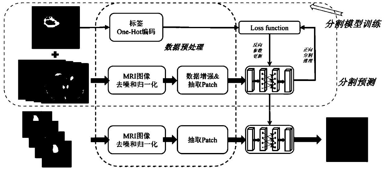
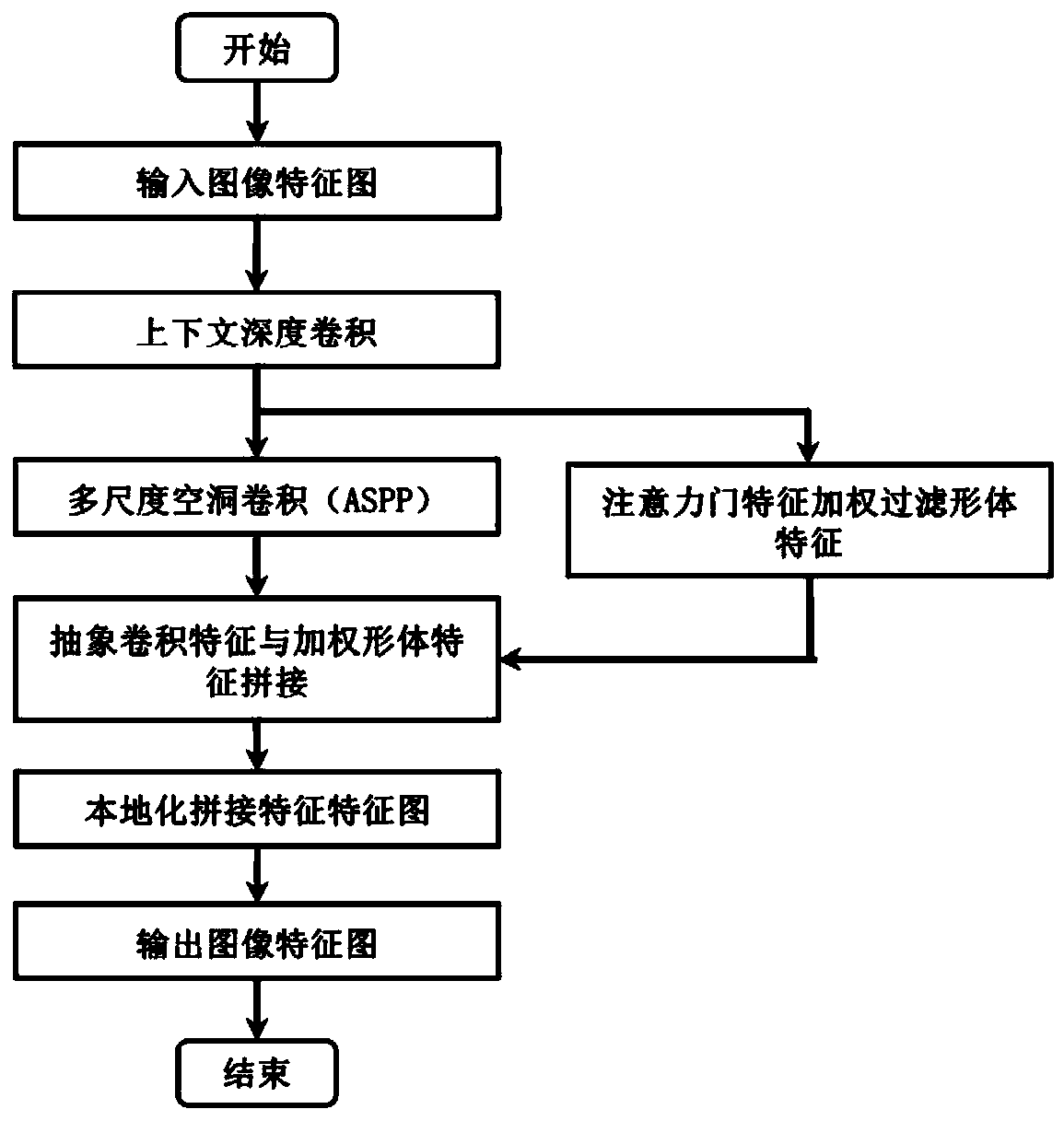
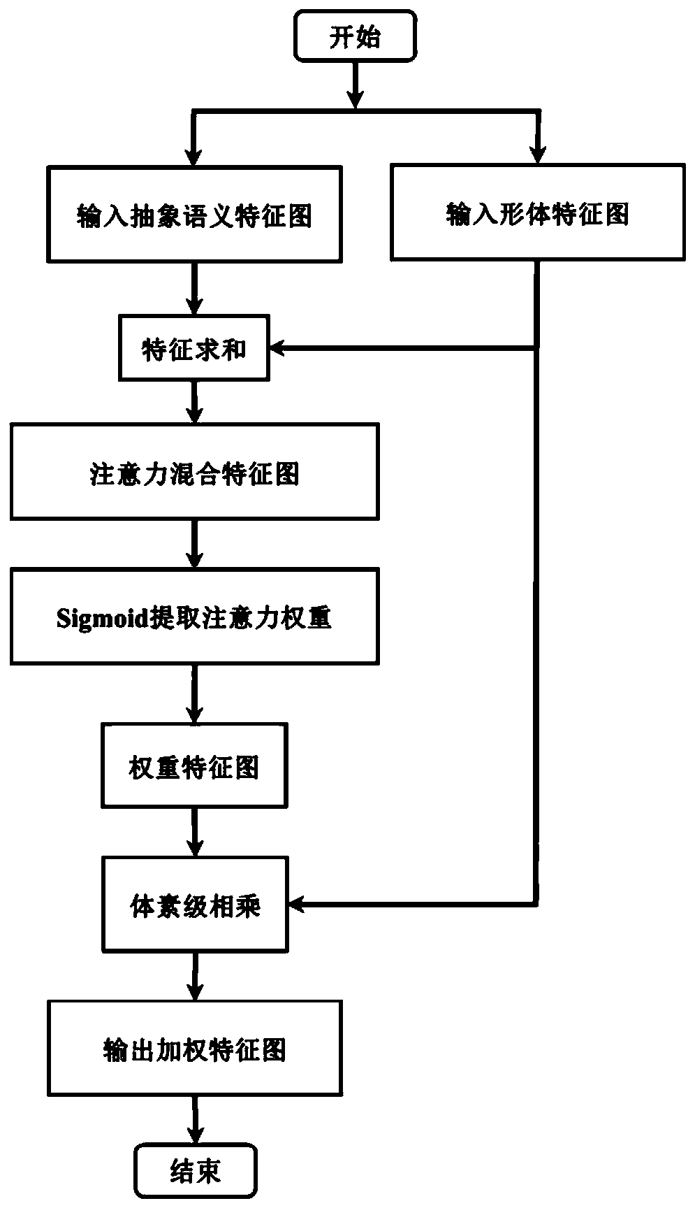
[0028] Such as Figure 1-Figure 3 In combination with the above, the present invention discloses a method for multi-modal brain glioma image segmentation with an adaptive attention gate with multi-level features. The method includes the following steps:

[0029] S1: Preprocessing the MRI glioma image data to obtain a data sample set;

[003...


PUM


Abstract

The invention discloses a multi-modal brain glioma image segmentation method of an adaptive attention gate. The method comprises the steps of S1, preprocessing the MRI glioma image data to obtain a data sample set; S2, building a segmentation network model of the adaptive attention gate with multi-level features, and training the segmentation model; S3, performing lesion segmentation prediction byusing the trained segmentation network model. According to the present invention, the attention gate can automatically learn a group of weights to represent the importance of each layer of features in the body features, the non-pathological noise of the superficial layer features is adaptively suppresses, and the lesion details are added to the deep layer features; a tumor segmentation network adopts a mixed loss function combining a binary cross entropy loss function and a dice loss function to train a model. According to the method, the semantic information and the detail features of different levels of a deep full convolutional network are fully mined, and under the action of the attention gate, the best glioma segmentation effect is achieved by fusing the features of different levels.

Description

technical field [0001] The present invention relates to a technology for glioma image segmentation based on a fully convolutional deep neural network, in particular to a multimodal glioma image segmentation method with an adaptive attention gate with multi-level features. Background technique [0002] Glioma has the highest mortality rate among brain tumors. These tumors can be divided into low-grade gliomas (LGG) and high-grade gliomas (HGG), with the former being less aggressive and aggressive than the latter. In clinical practice, further informative MRI sequences are particularly useful in the assessment of gliomas. Accurately segmenting gliomas and their internal structures is important not only for treatment planning, but also for subsequent evaluations. However, manual segmentation is time-consuming and subject to internal and internal scoring errors that are difficult to represent. For these reasons, precise semi-automatic or automatic methods are required. Howev...

Claims


Application Information

Patent Timeline
IPC IPC(8): G06T7/13G06T5/00
CPCG06T7/13G06T2207/10088G06T2207/30096G06T5/70
Inventor 郭顺杰曾卫明邓金石玉虎郭健
Owner SHANGHAI MARITIME UNIVERSITY
Learn More