Method for segmenting myopia macular lesion area in retina OCT image based on improved U-shaped network
A technology for macular degeneration and region segmentation, applied in image analysis, image enhancement, image data processing, etc., can solve problems such as poor performance of small target segmentation and lack of context information, achieve fast and effective reconstruction of high resolution and reduce information loss , Speed up the effect of network convergence
Active Publication Date: 2021-02-02
SUZHOU UNIV
View PDF9 Cites 0 Cited by
- Summary
- Abstract
- Description
- Claims
- Application Information
AI Technical Summary
Problems solved by technology
[0004]
This application aims to solve the above-mentioned technical problems, and provides a method for segmenting myopic maculopathy in retinal OCT images based on an improved U-shaped network, so as to realize the automatic segmentation of RBCC damage and myopic traction lines in retinal OCT images, and solve the problems in the prior art The problem of poor performance of small target segmentation caused by the lack of context information in the U-shaped deep learning network
Method used
View moreImage
Smart ImageViewing Examples

Examples
Embodiment
[0025] Embodiment: a method for segmenting the myopic maculopathy region based on the retinal OCT image of the improved U-shaped network, the method comprising:
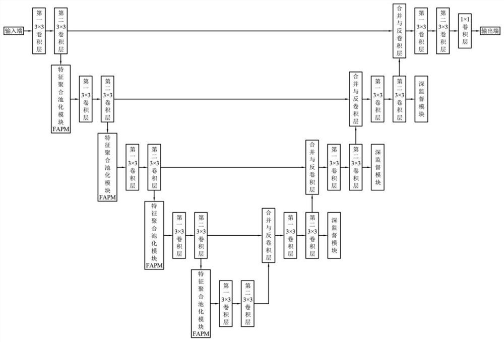
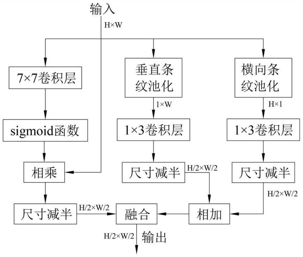
[0026] Build a network structure, which includes an encoder module, a feature aggregation pooling module FAPM, a decoder module, and a deep supervision module. The feature aggregation pooling module FAPM is set in the encoder channel of the encoder module for downsampling and aggregation. Context information and local information, the deep supervision module is set in layers other than the bottom layer of the decoder to guide the network to generate more representative feature maps;
Login to View More PUM
 Abstract
The invention discloses a method for segmenting a myopia macular lesion area in a retina OCT image based on an improved U-shaped network, and the method comprises the steps: building a network structure which comprises an encoder module, a feature aggregation pooling module FAPM, a decoder module and a deep supervision module, wherein the feature aggregation pooling module FAPM is arranged in an encoder channel of the encoder module and used for down-sampling and aggregating global context information and local information, and the deep supervision module is arranged in each layer, except a bottom layer, of the decoder and used for guiding a network to generate a feature map with higher representativeness, in an experiment, the output of the network structure is verified by adopting a joint segmentation task of RBCC damage and myopia traction streak lesion areas in an optical coherence tomography OCT image. According to the method for segmenting the myopia macular lesion area in the retina OCT image based on the improved U-shaped network, automatic segmentation of RBCC damage and myopia traction lines in the retina OCT image is achieved, and the small target segmentation performance is improved.
Description
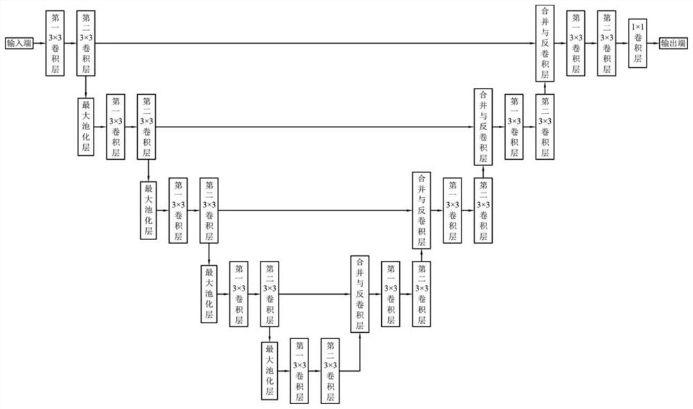
technical field [0001] The present application relates to the technical field of retinal image segmentation methods, in particular to a method for segmenting myopic maculopathy regions in retinal OCT images based on improved U-shaped networks. Background technique [0002] Currently the most commonly used medical segmentation network U-Net has a relatively simple structure, as shown in the accompanying drawings figure 2 As shown, the function of the encoder part on the left in the figure is feature extraction, and the part of the decoder on the right is used for upsampling. This structure is called an encoder-decoder structure. Because the overall structure of this network is similar to the uppercase English letter U, it is named U-Net. U-Net is very different from other common segmentation networks: U-net adopts a completely different feature fusion method, that is, through skip connections, features are merged together in the channel dimension in a splicing manner to form...
Claims
Login to View More Application Information
Patent Timeline
 IPC IPC(8): G06T7/11G06T3/40G06N3/04G06N3/08
CPCG06T7/11G06T3/4053G06T3/4046G06N3/084G06T2207/10101G06T2207/20081G06T2207/20084G06T2207/30041G06N3/045
Inventor 朱伟芳刁逸超陈新建
Owner SUZHOU UNIV