Automatic segmentation method for articular cartilage tissue in three-dimensional medical image

A medical image and articular cartilage technology, applied in the field of medical image processing, can solve the problems of less articular cartilage data sets and less segmentation methods, and achieve the effects of being easy to use, improving accuracy, and slowing down the speed of evolution

Inactive Publication Date: 2020-06-30
南京汇百图科技有限公司
View PDF4 Cites 2 Cited by
  • Summary
  • Abstract
  • Description
  • Claims
  • Application Information

AI Technical Summary

Problems solved by technology

[0003] Although some progress has been made in the segmentation of articular cartilage in 3D medical images, the following problems still exist: 1) Most of the proposed methods are applied to knee articular cartilage t...

Method used


Image

  • Automatic segmentation method for articular cartilage tissue in three-dimensional medical image
  • Automatic segmentation method for articular cartilage tissue in three-dimensional medical image
  • Automatic segmentation method for articular cartilage tissue in three-dimensional medical image

Examples


Embodiment Construction

[0027] The present invention will be described in further detail below in conjunction with the accompanying drawings and specific implementation processes.

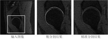
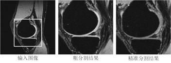
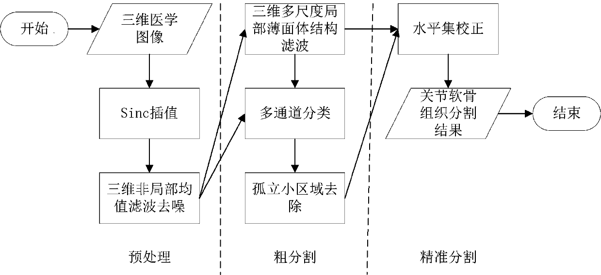
[0028] Such as figure 1 As shown, the present invention proposes an automatic segmentation method of articular cartilage tissue in three-dimensional medical images, which is divided into three steps: preprocessing, rough segmentation, and precise segmentation.

[0029] Step 1: Preprocessing. A 3D medical image composed of a sequence of N slice images is used as input data, and the preprocessing step includes two sub-steps of interpolation and denoising.

[0030] Step 1.1: Interpolation. Since the voxel spacing between slices in medical images is often greater than the voxel spacing within slices, medical images are non-isotropic (that is, voxel anisotropy), and the extraction of rotation-invariant features requires 3D medical image data to be isotropic. linear (i.e. voxels are isotropic). In order to obtain isotropic ...


PUM


Abstract

The invention provides an automatic segmentation method for articular cartilage tissue in a three-dimensional medical image. The automatic segmentation method is composed of three parts of three-dimensional medical image preprocessing, articular cartilage tissue coarse segmentation and articular cartilage tissue correction. According to the automatic segmentation method for the articular cartilagetissue in the three-dimensional medical image, the three-dimensional medical image is input, and the corresponding articular cartilage tissue is output. The method is an unsupervised segmentation method, can quickly and automatically realize cartilage tissue segmentation in a three-dimensional medical image without any manually labeled training sample, and has segmentation precision equivalent tothat of a supervised segmentation method.

Description

technical field [0001] The invention belongs to the technical field of medical image processing, and in particular relates to an automatic segmentation method of articular cartilage tissue in a three-dimensional medical image. Background technique [0002] Osteoarthritis is the most common joint disease in the world, mainly caused by lesions of articular cartilage tissue, and its incidence and prevalence increase with age. In addition to causing physical and psychological sequelae, it is also accompanied by a huge social and economic burden. Accurate segmentation of articular cartilage tissue in 3D medical images can assist doctors in analyzing the morphology of cartilage and provide quantitative data for determining the severity of osteoarthritis. [0003] Although some progress has been made in the segmentation of articular cartilage in 3D medical images, the following problems still exist: 1) Most of the proposed methods are applied to knee articular cartilage tissue, an...

Claims


Application Information

Patent Timeline
IPC IPC(8): G06T7/10G06T19/20G06T5/00
CPCG06T7/10G06T19/20G06T5/002G06T2207/10012G06T2207/30008
Inventor 常勇刘慧芳
Owner 南京汇百图科技有限公司
Why Eureka
Try Eureka