Aortic valve fast segmentation method based on esophageal echocardiography

A technology of aortic valve and ultrasound, applied in image analysis, image data processing, instruments, etc., can solve problems such as overflow and incomplete ultrasound image segmentation

Inactive Publication Date: 2013-05-08
HEBEI UNIVERSITY
View PDF5 Cites 10 Cited by
  • Summary
  • Abstract
  • Description
  • Claims
  • Application Information

AI Technical Summary

Problems solved by technology

[0004] The purpose of the present invention is to provide an automatic, fast and accurate method for segmenting the aortic valve in ultrasonic images, so as to solve the problems of incomplete and serious overflow in the ultrasonic image segmentation of the aortic valve in the prior art

Method used


Image

  • Aortic valve fast segmentation method based on esophageal echocardiography
  • Aortic valve fast segmentation method based on esophageal echocardiography
  • Aortic valve fast segmentation method based on esophageal echocardiography

Examples


Embodiment

[0046] The invention is Dual-Core CPU E5800 3.20GHz, graphics card is NVIDIA GeForce GT430NVIDIA GeForce GT430, memory is 2.00GB, operating system is WindowXP computer implementation, the whole segmentation method is written in C++ and Matlab language.

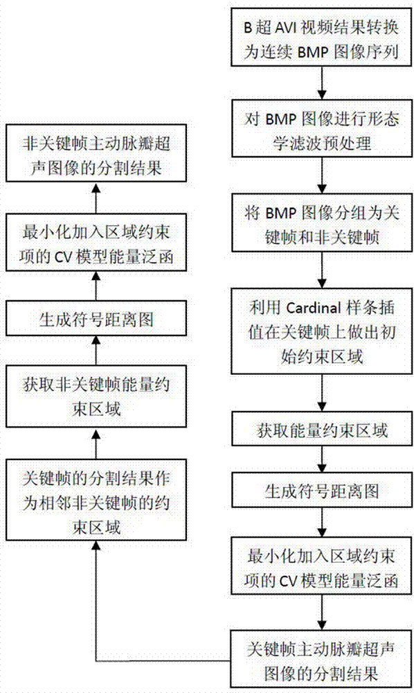
[0047] (1) The B-ultrasound video results (AVI format) output by transesophageal ultrasound use the DirectShow platform and the FFDShow video format decoder to convert the AVI format video files into 24-bit or 8-bit BMP format continuous ultrasound image sequences. Arrange the ultrasound image sequence in chronological order.

[0048] (2) Perform morphological filtering preprocessing on the above continuous image sequence: perform closed operation on the original image of the ultrasonic image to obtain the marked image, then perform erosion operation on the marked image and perform intersection operation with the original image until the iteration ends when convergence. The preprocessed ultrasound image can not only reduce t...


PUM


Abstract

The invention discloses an aortic valve fast segmentation method based on esophageal echocardiography. Due to the fact that a constraint area is defined on a critical frame of an ultrasound image, an area constraint term is produced by the constraint area, energy evolution coefficient of variation (CV) module is added, and energy functional of the CV module is minimized, an aortic valve segmentation result of the critical frame is obtained. Meanwhile, due to the fact that the segmentation result of the critical frame is used as a constraint area of a non-critical frame, the constraint area is added with the energy evolution CV module, and the CV module energy functional is minimized, and an aortic valve segmentation result of the non-critical frame is obtained. The aortic valve fast segmentation method based on the esophageal echocardiography has the advantages of being capable of fast and accurately segmenting an aortic valve of the ultrasound image.

Description

technical field [0001] The invention relates to an aortic valve segmentation method of an ultrasonic image, in particular to a rapid aortic valve segmentation method based on esophageal ultrasound. Background technique [0002] In modern medical imaging, ultrasound images have the advantages of low intensity, low price, and harmless to the human body, especially for the detection of soft tissues and the observation of blood flow in cardiovascular organs. With the improvement of living standards and the aging of the population, there are more and more common heart valve diseases such as aortic valve stenosis, aortic valve insufficiency and aortic valve prolapse. Clinically, the main diagnostic method for such diseases is to use ultrasound equipment to observe the shape and movement of the valve. Echocardiography is a good tool for detecting heart valve diseases. The first step in its analysis is ultrasonic medical image segmentation. Because there are many speckle noises in ...

Claims


Application Information

Patent Timeline
Patent Type & Authority Applications(China)
IPC IPC(8): G06T7/00
Inventor 顾力栩董斌郭怡婷王兵
Owner HEBEI UNIVERSITY
Why Eureka
Try Eureka